
Figure 2.
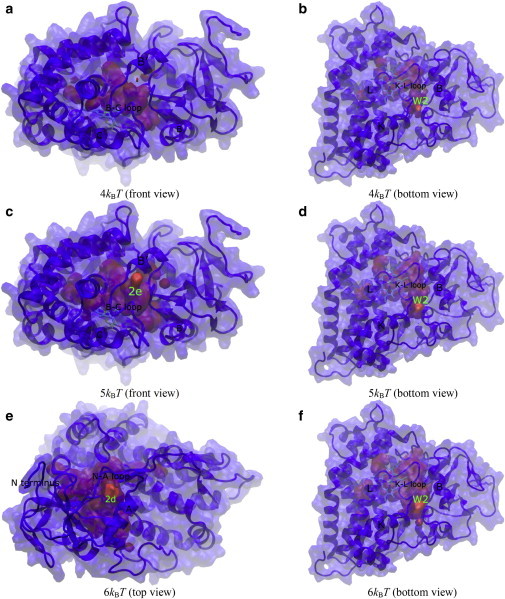
Free-energy isosurfaces of hydration in apo-P450cam. Relevant protein secondary structures and water diffusion exits on the protein surface are labeled. Front, top, and bottom views of the protein are looking down the I helix, looking down on the heme plane from the distal side, and looking up from below the heme plane, respectively.