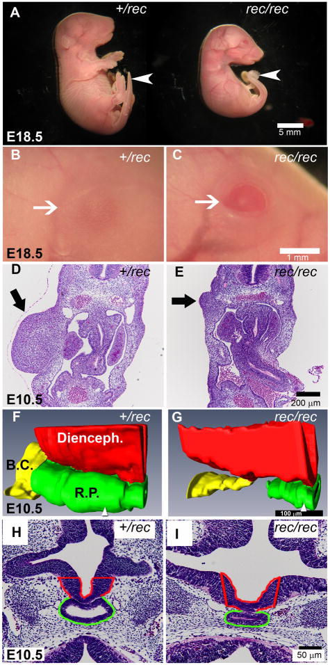
Figure 2. Morphology of Fgfr2IIIbrec/rec mutants.
(A) Low magnification views of Fgfr2IIIbrec/+ and Fgfr2IIIbrec/rec embryos. At E18.5, the Fgfr2IIIbrec/rec mutant lacks limbs, is smaller than its heterozygous littermate, and also has a curved tail (white arrow). (B and C) Higher magnification of the eye (white arrow) from embryos shown in (A). The Fgfr2IIIbrec/rec embryo has an eyelid defect. (D and E) Transverse sections through the limb region at E10.5. The Fgfr2IIIbrec/rec mutant (E, black arrow) exhibits defects in the limb mesenchyme. (F–I) Pituitary development at E 10.5. (F and G) 3-dimensional reconstructions of Rathke’s pouch (RP), the Buccal cavity (BC), and the diencephalon (Di) generated from transverse sections. (H and I) Representative sections from the specimens reconstructed in F and G. Position of the sections (H and I) within each reconstruction are indicated by the white arrowhead at the bottom of panels F and G. In the Fgfr2IIIbrec/rec mutant, Rathke’s pouch is narrower in the dorsal-ventral axis and no longer continuous with the Buccal cavity.

