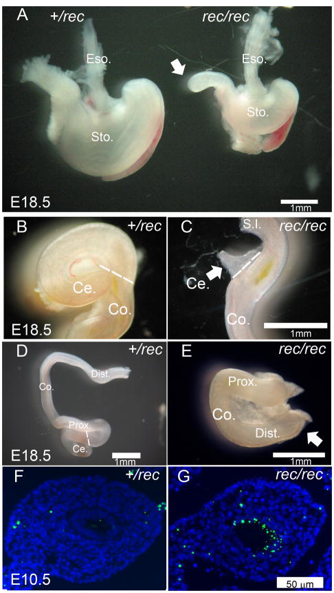
Figure 4. Alimentary track development in Fgfr2IIIbrec/rec mutants.
(A) Stomach and duodenum of Fgfr2IIIbrec/+ and Fgfr2IIIbrec/rec embryos at E18.5 (B and C) the cecum, (D and E) and the colon. White arrows in A and E indicate atresias. White arrow in C indicates malformed cecum. Broken white line in B, C, and D indicates boundary between cecum and colon. TUNEL staining of transverse sections of colon in (F) Fgfr2IIIbrec/+ and (G) Fgfr2IIIbrec/rec embryos at E10.5.

