
Figure 3. Sox3 expression is lost in tcf7l1 morphants.
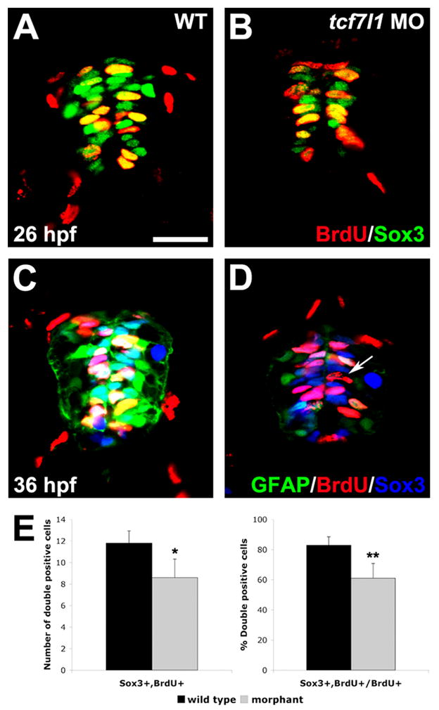
(A,B) BrdU and Sox3 co-immunohistochemistry in spinal cross-sections of wild-type and tcf7l1 morphant embryos. Sox3 expression is reduced in tcf7l1 morphants at 26 hpf. (C,D) BrdU, Sox3 and gfap:GFP triple-labeling at 36 hpf. Expression of Sox3 and GFAP are greatly reduced in tcf7l1 morphants (D) compared to wild-type embryos (C). Arrow indicates a BrdU+ cell without expression of GFAP or Sox3. (E) Number and percent of BrdU/Sox3 double-positive cells/section in wild-type and tcf7l1 morphant spinal cords. Error bars indicate SD and asterisks indicate statistical significance. *p < 0.05, **p<.0.001 by Student’s unpaired t-test. n=12 sections from 5 individual embryos for each bar. Scale bar = 20μM.