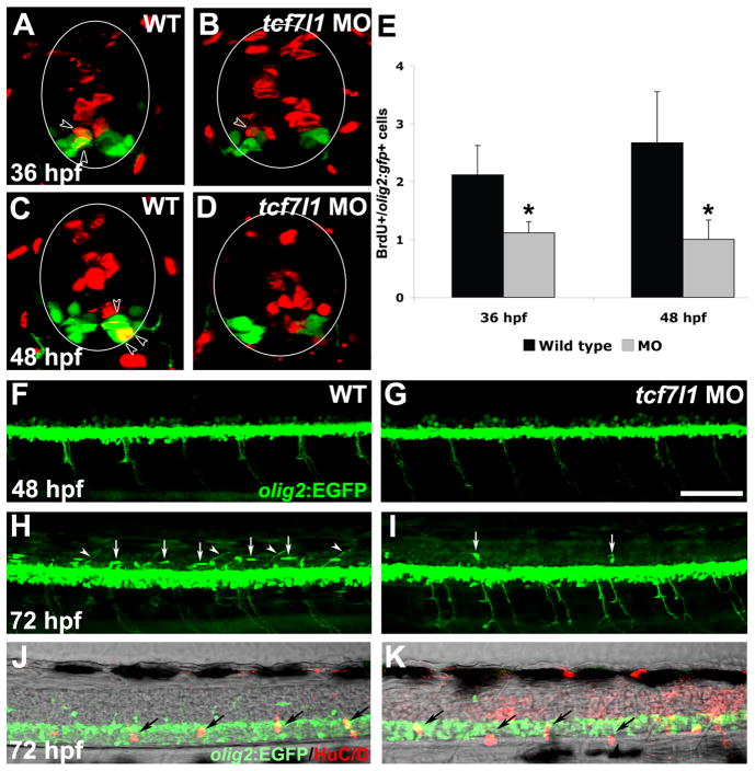
Figure 5. OPC development is reduced in tcf7l1 morphants.
(A–D) BrdU and GFP double immunohistochemistry on cryosections of wild-type and tcf7l1 morphant Tg(olig2:EGFP)vu12 embryos. Spinal cord is outlined in circles and arrowheads indicate double-labeled cells. In tcf7l1 morphants, fewer BrdU+ cells are observed within the GFP+ population. (G) Number of BrdU+/olig2:gfp+ cells per cryosection in wild-type embryos and tcf7l1 morphants at 36 and 48 hpf. Error bars indicate SD and asterisks indicate statistical significance. *p < 0.05 by Student’s unpaired t-test. n=9 sections from 3 different embryos for each bar. (F–K) Confocal projection images showing lateral views of the spinal cord in Tg(olig2:EGFP)vu12 embryos. (F,G) GFP-positive cells are reduced in tcf7l1 morphants at 48 hpf. (H,I) At 72 hpf, dorsally migrating OPCs (arrows) and dorsally projecting motoneurons (arrowheads) are absent in tcf7l1 morphants. (J,K) The decrease in OPCs is not due to a general developmental delay, as normal dorsal root ganglion development (arrows) is observed in tcf7l1 morphants. Scale bars = 20μM in D, 40μM in F.

