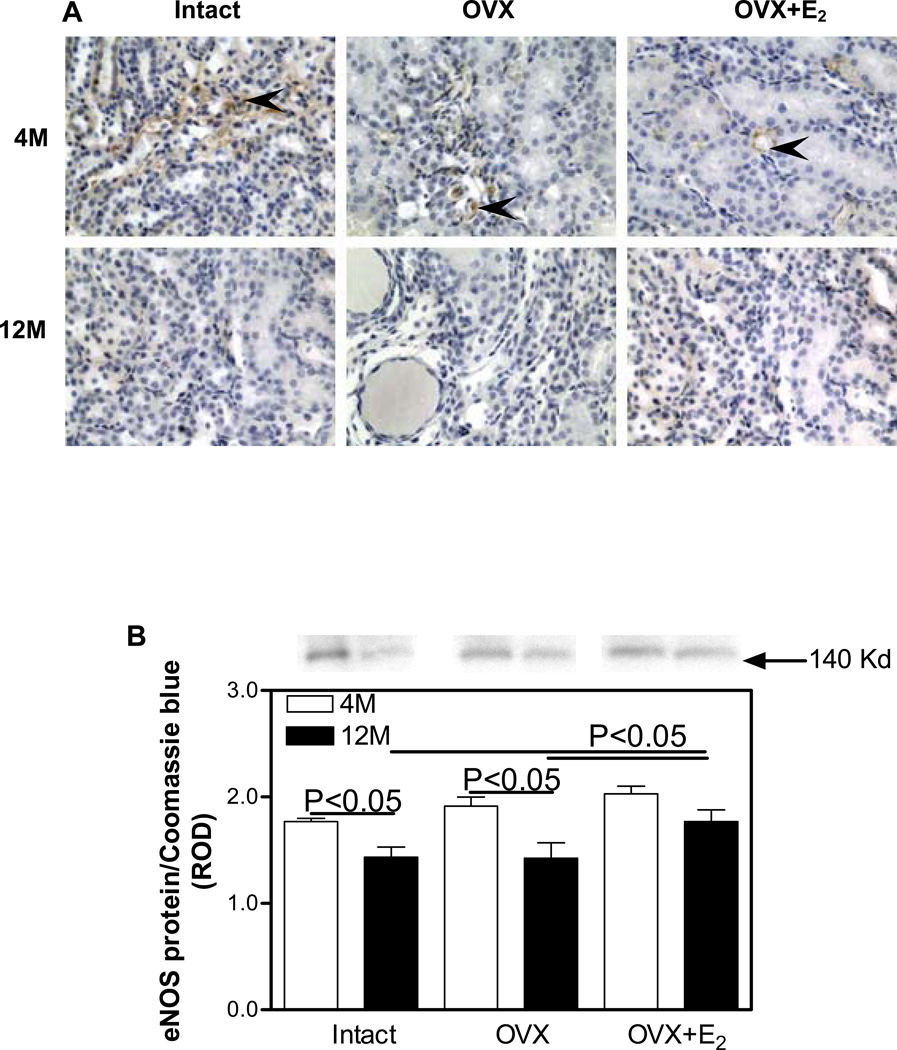
Figure 4.
Immunohistochemical localization and expression of eNOS protein in the renal medulla. A. eNOS (brown staining) immunolocalization. Arrow heads, endothelial cells. Original magnification ×400. B. eNOS protein expression by Western blotting. Top panel, representative gels of eNOS protein expression. Bottom panel, densitometric scans of eNOS protein levels in relative optical density (ROD) expressed as a ratio of eNOS/Coomassie blue. Data are expressed as mean±SEM.

