
Figure 4.
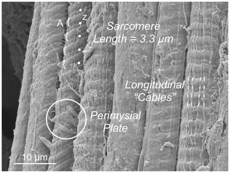
Scanning electron micrograph of 7 adjacent muscle fibers. Note that the surface topology varies among the fibers. In 5 cases, surface striations are visible, some of which clearly show A-band (curved brackets) and Z-band periodicity (dots). In 3 fibers, stout longitudinal “cables” extend a distance of at least 100 μm. In one case, a discrete connection is seen on the surface of a fiber that presumably represents the “perimysial plate” (circled) that connects adjacent fibers. Micrograph was obtained from a mouse EDL muscle stretched to a sarcomere length of 3.3 μm.