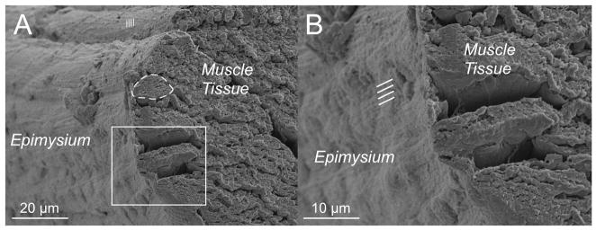
Figure 7.
Epimysial layer of a mouse EDL muscle viewed in cross-section. A connective tissue sheath is clearly seen surrounding muscle fibers. (A) Survey view of the muscle where a region can be observed with sarcomere periodicity through the connective tissue layer. An individual muscle fiber is outlined (dashed line) (B) Closer view of the epimysium reveals longitudinal periodicities (lines) with approximately 1 μm of spacing.

