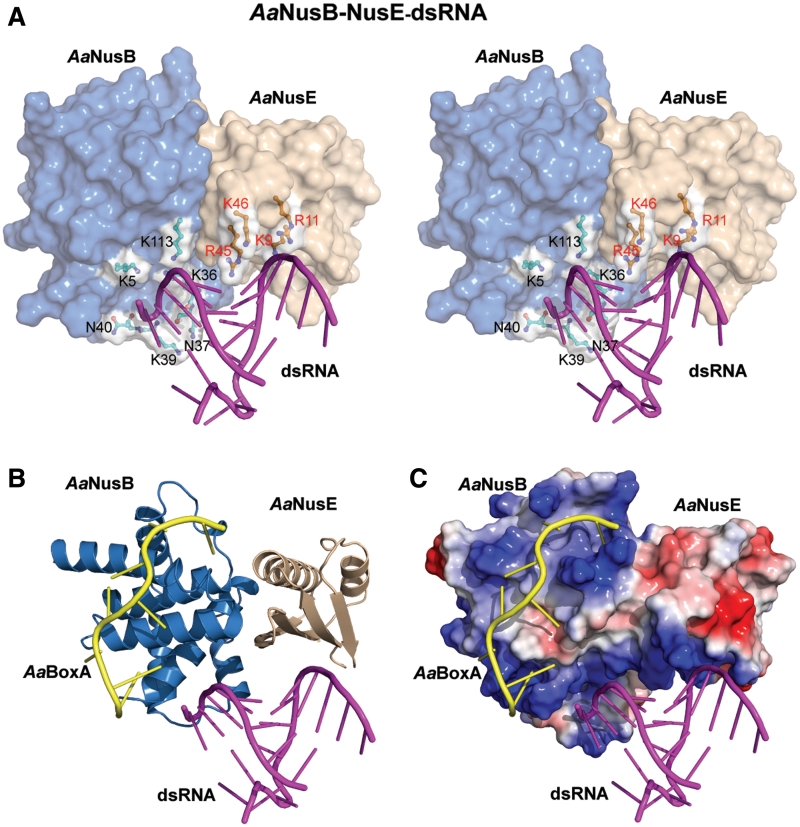
Figure 5.
Binding of dsRNA by the NusB–NusE heterodimer. AaNusB molecules are colored in blue, AaNusE molecules in wheat and dsRNA in magenta. Orientations in all panels are identical to that in Figure 2A. (A) Crystal structure of the AaNusB–NusE–dsRNA ternary complex in stereo. Residues involved in dsRNA binding are depicted as ball-and-sticks with white surface. (B and C) Superposition of the crystal structures of AaNusB–NusE–BoxA and AaNusB–NusE–dsRNA, illustrating the contiguous binding regions for AaBoxA and dsRNA. In panel C, the protein surface representation is colored by vacuum-electrostatic potential (red, negative; blue, positive), which depicts the AaBoxA and dsRNA binding sites as a contiguous positively-charged surface.

