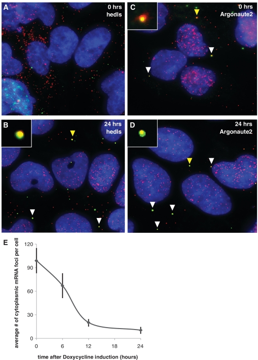
Figure 2.
Single molecule FISH combined with immunofluorescence shows mRNA co-localization with RISC and P body proteins when miRNA is expressed. (A–D) smFISH-IF co-staining of protein antibody (green, Cy-3) and luciferase reporter mRNA (red, Cy-5) at various times after doxycycline induction of mRNA. Blue = DAPI. White arrows point to examples of co-localization. Yellow arrows point to co-localization shown at 5× size in the inset of the same picture. (A) Hedls, 0 h. (B) Hedls, 24 h. (C) Argonaute, 0 h. (D) Argonaute2, 24 h. (E) Luciferase reporter mRNA levels are reduced when miRNA is expressed. Average number of cytoplasmic mRNA foci per cell 0, 6, 12 and 24 h after doxycycline induction of miRNA. N = 52 cells for 0 h, 48 cells for 6 h, 51 cells for 12 h and 50 cells for 24 h. Error bars = 95% confidence interval for means.

