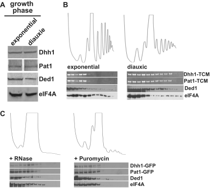
Figure 3.
Analysis of Dhh1, Pat1, eIF4A and Ded1 co-sedimentation across polysomal gradients. (A) The same number of cells in glucose-fermenting exponential phase or post-diauxic shift phase were extracted and proteins resolved by SDS–PAGE and western blotting to determine relative protein expression levels for the proteins indicated. (B) Polysomal gradient profiles and corresponding fractions were generated with either exponentially growing or post-diauxic shift cell cultures expressing either Dhh1-TCM or Pat1-TCM and protein distributions were determined by in-gel fluorescence (Dhh1-TCM and Pat1-TCM) or by western blotting (Ded1 and eIF4A). (C) Post-diauxic shift cell cultures expressing either Dhh1-GFP or Pat1-GFP were treated with RNase A or puromycin and polysomal gradient profiles and corresponding fractions were collected. Dhh1 and Pat1 distributions were determined using anti-GFP antibodies while Ded1 and eIF4A were identified using protein-specific polyclonal antibodies.

