Figure 4.
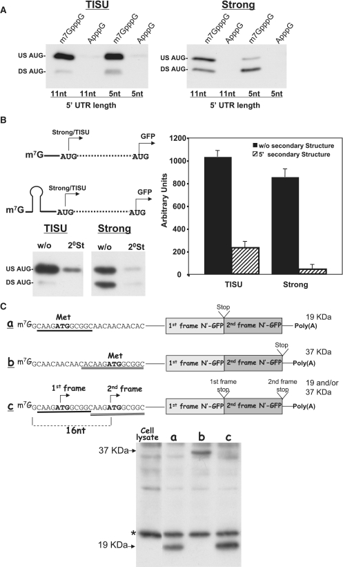
Translation initiation from TISU is dependent on 7mG cap. (A) mRNAs bearing AUG with either TISU or a strong contexts located either 5 or 11 nt from the 5′-termini were in vitro transcribed in the presence of m7GpppG cap or the unmethylated cap-analog ApppG. The mRNAs were translated in vivo by transfection into 293T cells. Cell lysate was prepared 24 hours following transfection and subjected to western blot using anti-GFP. Translation efficiency is shown in the representative blot of three independent experiments. (B) The effect of AUG upstream secondary structure on the efficiency and fidelity of translation from a strong and TISU AUG contexts. The upper-left panel shows a schematic representation of a secondary structure located upstream from the AUG within TISU or strong AUG context. The constructs with or without secondary structure were in vitro transcribed and then translated in vivo by transfection into 293T cells and translation efficiency was assayed by blotting with anti-GFP. Representative immunoblot for the strong and the TISU AUG contexts are shown in the bottom-left panel and the graphs represent the average ± SD of the intensity of the upstream (30 KDa) translation site of four independent experiments. (C) Recognition of TISU's AUG is dependent on the 5′-end of mRNA. The upper panel shows a schematic representation of mRNAs bearing either one or two TISU elements in tandem with the expected protein size translated from each AUG. The mRNAs were transcribed and capped in vitro and then were translated in vivo by transfection into 293T cells. Cell lysate was prepared 24 hours following tranfection and subjected to western blot using anti-GFP (lower panel). The positions of 19 and 37 KDa proteins are shown by arrows.

