Figure 7.
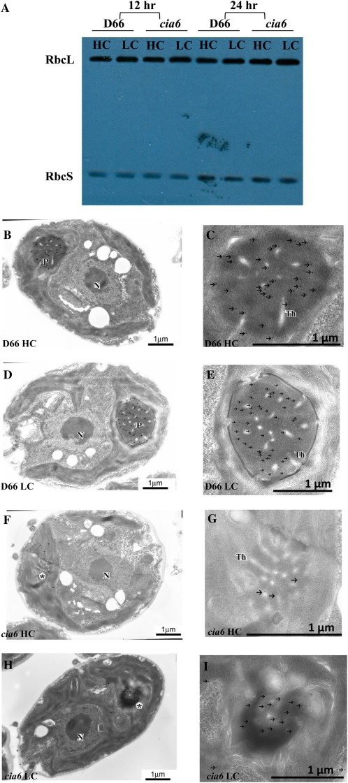
Rubisco content in the wild-type D66 and mutant cia6 cells was investigated using western blotting (A) and immunogold labeling (B–I) against anti-Rubisco antibody. A, Total protein isolated from D66 and cia6 grown under high- and low-CO2 conditions (HC and LC, respectively) for 12 and 24 h. Using western blotting, the amount of dissociated Rubisco large and small subunits (RbcL and RbcS) was estimated to be normal in the mutant compared with the wild type. B and C, High-CO2-grown wild-type D66 cells grown on minimal medium probed with an antibody raised against Rubisco. D and E, Low-CO2-grown wild-type D66 cells grown on minimal medium probed with an antibody raised against Rubisco. F and G, High-CO2-grown cia6 cells grown on minimal medium probed with an antibody raised against Rubisco. H and I, Low-CO2-grown cia6 cells grown on minimal medium probed with an antibody raised against Rubisco. N, Nucleus; P, pyrenoid; Th, thylakoid. Asterisks indicate pyrenoid-like structures in cia6. Bars = 1 μm. [See online article for color version of this figure.]

