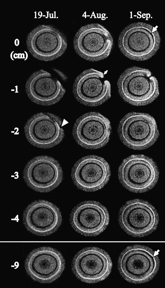
Figure 2.
Cross-sectional MR images acquired from interval positions of 1 cm pitch within the monitoring region of the 1-year-old stem of the control tree after injection with distilled water. MRI parameters were as follows: repetition time = 500 ms, echo time = 19 ms, slice thickness in the axial direction = 1 mm. The arrowhead indicates injury by knife. The small arrow indicates newly made tissue that originated in the cambium. The large arrows indicate the embolism that was made within latewood of the injection site in the current year.

