Figure 5.
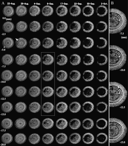
Cross-sectional MR images obtained from interval positions of 2.5 mm pitch within the monitoring region of a 1-year-old stem of Ino-2 after inoculation with PWN. MRI parameters were as follows: repetition time = 500 ms, echo time = 19 ms, slice thickness in the axial direction = 1 mm. A, Global development of xylem dysfunction in the monitoring region. The arrowhead indicates the embolism that was made within latewood in the current year. The arrow indicates the starting region of the massed embolism. The dotted grid denotes the extensional region in B. B, Magnification of the right half of the MR images between –7.5 and –15.0 mm on September 8. The scattered embolisms were two different types: longitudinal (arrowheads) and radial (arrow).

