
Figure 12.
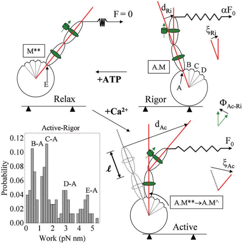
Myosin cross-bridge under rigor, relaxed, and active isometric conditions. The cross-bridge consists of an actin-binding globular domain and lever-arm with two light chains. GFP and its dipole moment are depicted as a green cylinder and arrow adjacent to RLC. Cross-bridges in rigor have different RLC-GFP orientations due to lever-arm deformation induced by the lattice force αF0 where α is a constant less than 1 and F0 is active isometric tension. The binding of ATP to myosin in rigor relaxes the cross-bridge, relieving shear strain and repriming the lever-arm. Addition of Ca2+ to the relaxed cross-bridge activates actin binding for active tension and shear strain development. tan ξ = d/l, for d the deformation length and l the original length of the deformed member, is the shear strain in active or rigor cross-bridges. The work histogram is computed using eq 4 and the active–rigor transitions with cos(ξAc − ξRi) = cos(ΦAc—Ri). The work histogram indicates substates in the power-stroke that we model as a series of attitudes labeled A–E that the lever-arm can assume during its swinging motion. Some of the biochemical states from Scheme 1 are assigned to the lever-arm attitudes including rigor (A.M, position A), relax after repriming (M**), or active (A.M**) (position E). Active state contains a series of substates (E–B) that upon transition to rigor give discretized peaks in the work histogram. The B → A transition is depicted in the figure but C → A, D → A, and E → A are also represented in the work histogram.