Abstract
Wnt signaling pathways control lineage specification in vertebrate embryos and regulate pluripotency in embryonic stem (ES) cells, but how the balance between progenitor self-renewal and differentiation is achieved during axis specification and tissue patterning remains highly controversial. The context- and stage-specific effects of the different Wnt pathways produce complex and sometimes opposite outcomes that help to generate embryonic cell diversity. Although the results of recent studies of the Wnt/β-catenin pathway in ES cells appear to be surprising and controversial, they converge on the same conserved mechanism that leads to the inactivation of TCF3-mediated repression.
Keywords: Embryonic stem cells, TCF, β-catenin, GSK3, Pluripotency, Self-renewal, Homeodomain-interacting protein kinase
Introduction
A major goal of developmental and stem cell biology is to elucidate the mechanisms that allow embryonic progenitor cells to choose a specific path for differentiation or to maintain their pluripotency. Pluripotency is a common attribute of the early blastomeres of vertebrates, and is one that allows these cells to contribute to any of the three germ layers (ectoderm, mesoderm or endoderm). The derivation of pluripotent and self-renewing embryonic stem (ES) cells in the early 1980s established one of the best in vitro models of early embryonic development (Evans and Kaufman, 1981; Martin, 1981). Nevertheless, our understanding of the key pathways that lead to the ES cell-like state is far from complete, mainly due to the fact that normal blastocyst cells do not undergo the unlimited self-renewal that is observed in ES cell culture. The question of how a blastocyst cell is able to differentiate into a cell that belongs to any of the three germ layers remains unclear. However, recent progress has provided some insight into this question, with the identification of several signaling pathways that are crucial for lineage specification in the early embryo and in ES cells (Arnold and Robertson, 2009; Boyer et al., 2005; Nichols and Smith, 2011; Rossant and Tam, 2009; Schier and Talbot, 2005; Ying et al., 2008).
Another critically important process in the embryo is the acquisition by cells of positional information. In the simplest case, this information corresponds to the specification of axes in the whole embryo and in specific organs. Proper positional values are essential for normal embryogenesis and need to be reconstituted during regeneration for proper organ function (Brockes and Kumar, 2008; Kragl et al., 2009). Whereas animal pole blastomeres of the Xenopus embryo appear to `remember' their position in the embryo from which they originated (Savage and Phillips, 1989; Sokol and Melton, 1991), there is no clear evidence that such positional information exists within the mammalian blastocyst from which ES cells are derived (Arnold and Robertson, 2009; Gardner et al., 1992; Rossant and Tam, 2009). Nevertheless, some studies argue that such information can arise by a stochastic mechanism de novo, during formation of ES cell aggregates called embryoid bodies (ten Berge et al., 2008b). Other experiments indicate that cell-cell signaling mediated in vivo by secreted molecules endows cells with positional information, which can be reconstituted to a limited degree in vitro, in embryoid bodies (ten Berge et al., 2008b).
One of the main signaling pathways that functions in the early embryo is the Wnt pathway, which is employed repeatedly during development and fulfils multiple roles (Clevers, 2006; van Amerongen and Nusse, 2009). Not only does Wnt signaling specify the anteroposterior (AP) body axis in most metazoan animals, but it has also been reported to promote ES cell pluripotency (Nusse et al., 2008; Wend et al., 2010), to specify the mesendodermal lineage and to inhibit neuroectodermal differentiation in mouse ES cells (Aubert et al., 2002; Bakre et al., 2007; Haegele et al., 2003; Lindsley et al., 2006; Sato et al., 2004) and in vertebrate embryos (Yoshikawa et al., 1997; Itoh and Sokol, 1999). Strikingly, whether Wnt ligands and receptors themselves have a proven role in pluripotency continues to be the subject of ongoing debate (Nusse et al., 2008; Wend et al., 2010). Although the major molecular players of the Wnt pathway are conserved, the mechanisms that endow this signaling pathway with stage-specific and cell context-dependent outcomes often remain unclear (Hoppler and Kavanagh, 2007; MacDonald et al., 2009; van Amerongen and Nusse, 2009). Further complexity has come with the realization that the individual components of this pathway have both Wnt-dependent and Wnt-independent functions. For example, glycogen synthase kinase 3 (GSK3), a central player in Wnt signaling, is also known to phosphorylate many cellular substrates and to modulate several pathways unrelated to Wnt (MacDonald et al., 2009). Thus, until a specific mechanism is unraveled, it remains formally possible that any of the Wnt pathway components could function to control ES cell pluripotency in a Wnt-independent manner.
In this review, I discuss the roles of Wnt proteins and the downstream components of the pathway, in particular β-catenin and T-cell factors (TCFs), in maintaining progenitor pluripotency and in allowing specific lineage decisions to be made in both ES cells and vertebrate embryos. Conclusions drawn from studies of Xenopus and mouse embryos highlight the existing controversies in the ES cell field and provide further insight into context-dependent TCF signaling mechanisms, which are likely to operate in all vertebrates. Although Wnt signaling has also been implicated in many morphogenetic processes, this subject has been extensively reviewed elsewhere (e.g. Angers and Moon, 2009; van Amerongen and Nusse, 2009; Wallingford et al., 2002), and as such will not be covered in this review.
The Wnt pathway in axis and germ layer specification
The main body plan of all vertebrate embryos is similar and involves the specification of the dorsoventral (DV) and AP axes and the proper positioning of the three germ layers (ectoderm, mesoderm and endoderm) during gastrulation. This is achieved primarily by cell-cell interactions mediated by the bone morphogenetic protein (BMP), fibroblast growth factor (FGF), Nodal and Wnt pathways, which constitute the major embryonic signaling pathways, the precise functions of which are still under investigation (Arnold and Robertson, 2009; Conlon et al., 1994; Harland and Gerhart, 1997; Rossant and Tam, 2009; Schier and Talbot, 2005; Sokol, 1999). Wnt signaling is widely utilized during early development to regulate body axis specification, germ layer formation and organogenesis (Clevers, 2006; van Amerongen and Nusse, 2009) (Fig. 1). The Wnt pathway also regulates the self-renewal of ES cells, one of the best in vitro models for studying pluripotency and lineage commitment (Nusse et al., 2008; Wend et al., 2010).
Fig. 1.
Wnt signaling regulates vertebrate body axis and germ layer formation. (A) Schematic of a Xenopus embryo at stage 10 (early gastrula). The animal pole (future anterior) is at the top and dorsal is to the right. During gastrulation, Wnt signaling antagonizes anterior neuroectoderm (ANE) development, but promotes posterior mesendoderm (PME, green) in the presence of Nodal-like signals. The dashed line indicates the prospective boundary between the ectoderm (orange) and the mesendoderm (yellow). The arrow indicates the direction of mesodermal cell movement towards the anterior end of the embryo during gastrulation. (B) The Wnt signaling gradient aligns with the anteroposterior (AP) embryonic axis. A stage 39 Xenopus tadpole, with posterior (P) to the left, anterior (A) to the right. Wnt signaling activity is graded from high (green) at the posterior to low (red) at the anterior. (C) Schematic of a mouse embryo at the primitive streak (PS) stages (E6.0-E7.0). The AP axis is indicated (left to right); the distal pole is at the bottom. The PS forms in a Wnt3-dependent manner at the posterior of the embryo and serves as the site of nascent mesoderm and definitive endoderm formation. The arrow indicates the direction of PS expansion towards the future anterior of the embryo. (A-C) Green dots represent nuclei with enriched β-catenin; green shading reflects high levels of Wnt signaling.
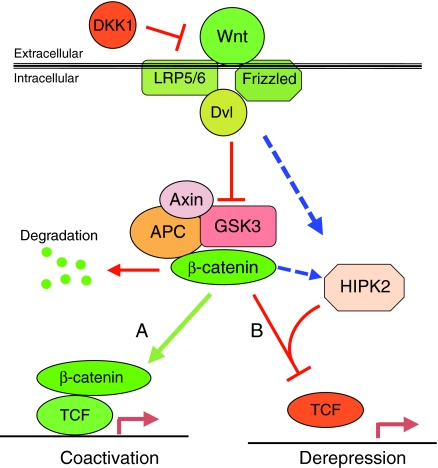
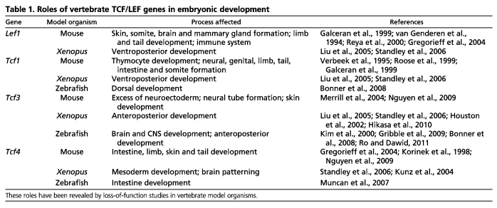
According to the commonly accepted scheme of the canonical Wnt signaling pathway, β-catenin is rapidly degraded by the complex of Axin, adenomatous (or adenomatosis) polyposis coli (APC) and GSK3 in the absence of a Wnt signal (Fig. 2). Wnt proteins act through various Frizzled receptors and LRP5/6 co-receptors to trigger downstream events, leading to the inactivation of this β-catenin-degradation complex. Stabilized β-catenin associates with transcription factors of the TCF/lymphoid enhancer factor 1 (LEF1) family, and this binding is thought to be essential for target gene activation (Arce et al., 2006; Archbold et al., 2011; Clevers, 2006). TCF proteins are the most distal components of the Wnt/β-catenin signaling cascade; they directly interact with target promoter DNA and as such are a key regulatory point in the pathway. The traditional view of this pathway has been that TCF proteins repress gene targets in the absence of the Wnt signal, but upon interaction with β-catenin are converted into transcriptional activators (Daniels and Weis, 2005; van de Wetering et al., 1997). In contrast to TCF proteins in Drosophila and C. elegans, which are represented by single genes, the four existing vertebrate TCF family members [TCF1 (also known as TCF7), LEF1, TCF3 (also known as TCF7L1) and TCF4 (also known as TCF7L2)] have acquired divergent functions, as revealed in depletion analyses (Liu et al., 2005; Standley et al., 2006) and in genetic double knockouts (Galceran et al., 1999; Gregorieff et al., 2004; Nguyen et al., 2009) (Table 1).
Fig. 2.
Schematic of the canonical vertebrate Wnt signaling pathway. Wnt proteins stimulate signaling via the Frizzled receptors, the LRP5/6 co-receptor and the Dishevelled protein to inactivate the β-catenin degradation complex, which consists of Axin, APC and GSK3. DKK1 is a secreted Wnt antagonist. (A) The inhibition of GSK3 activity results in β-catenin stabilization (green arrow) and in the transcriptional activation of target genes mediated by β-catenin in complex with TCF proteins. (B) Wnt signaling also stimulates the HIPK2-dependent elimination of TCF3-mediated target repression, during which β-catenin appears to act as a scaffold for TCF3 phosphorylation. Negative interactions are shown in red; dashed lines represent a hypothetical effect. APC, adenomatous polyposis coli; DKK1, dickkopf 1; Dvl, Dishevelled; GSK3, glycogen synthase kinase 3; HIPK2, homeodomain-interacting protein kinase 2; LRP5/6, low density lipoprotein receptor-related protein 5/6; TCF, T-cell factor.
Table 1.
Roles of vertebrate TCF/LEF genes in embryonic development
Previous studies have established an important role for Wnt/β-catenin signaling in the regulation of vertebrate axis and germ layer specification. A requirement for β-catenin in AP axis formation has been demonstrated in Xenopus, zebrafish and mouse embryos (Grigoryan et al., 2008; Heasman et al., 1994; Schier and Talbot, 2005). In mouse embryos that lack the Wnt3 (Liu et al., 1999), Lrp5/6 (Kelly et al., 2004) and β-catenin (Ctnnb1) (Haegel et al., 1995; Huelsken et al., 2000) genes, the body axis does not form properly and anterior neuroectoderm develops in excess. Conversely, the genetic inactivation of the negative regulators of the pathway, Axin1 (Zeng et al., 1997), Tcf3 (Kim et al., 2000; Merrill et al., 2004) or dickkopf 1 (Dkk1) (Mukhopadhyay et al., 2001), results in the development of ectopic axial structures and in posteriorization of the embryo, leading to microcephaly or the absence of the head. These studies highlight that Wnt signaling specifies the posterior region of the embryo, whereas inhibition of Wnt signaling is needed to specify the anterior. Studies in other animals, such as planarians, have revealed that this role of the Wnt/β-catenin pathway in AP axis specification is conserved in most metazoans (Petersen and Reddien, 2009). Whether and how this known function of Wnt signaling in embryonic development relates to the regulation of pluripotency and lineage decisions in ES cells remain important questions, which are discussed below.
Wnt signaling in ES cell pluripotency and lineage specification
ES cells are derived from the inner cell mass (ICM) of the mammalian blastocyst and represent a unique self-renewing embryonic progenitor population that has been `frozen' in its pluripotent state. ICM cells are also pluripotent, but quickly lose this property by committing to one of the three germ layers during gastrulation. The ability of mouse ES cells to self-renew has been attributed to the protein regulatory network that includes the pluripotency factors NANOG, OCT4 (also known as OCT3/4 or POU5F1 – Mouse Genome Informatics) and SOX2 (Boyer et al., 2005; Chambers et al., 2003; Jaenisch and Young, 2008; Loh et al., 2006; Mitsui et al., 2003; Niwa et al., 2000; Rosner et al., 1990; van den Berg et al., 2010). By contrast, human ES cells differ significantly from mouse ES cells in their required culture conditions (for which FGF2, but not LIF, is required) and contain lower levels of the pluripotency markers KLF4 and REX1 (ZFP42 – Human Genome Nomenclature Committee) (Nichols and Smith, 2011). Human ES cells are similar to mouse epiblast-derived stem cells (EpiSCs), which have a narrower developmental potential than ES cells, in support of the idea that they are captured at a later stage of development (Nichols and Smith, 2011).
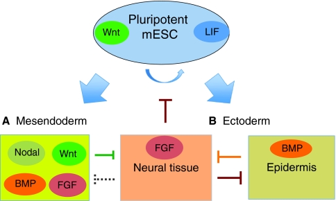
In addition to its known function in the early vertebrate embryo, Wnt signaling has been proposed to maintain the self-renewal and pluripotency of mouse ES cells (Anton et al., 2007; Hao et al., 2006; Kielman et al., 2002; Miyabayashi et al., 2007; Ogawa et al., 2006; Singla et al., 2006; ten Berge et al., 2011) (Fig. 3). Additionally, the Wnt pathway increases the efficiency with which differentiated cells can be reprogrammed toward pluripotency (Lluis et al., 2008; Marson et al., 2008). By contrast, the roles of the Wnt pathway in human ES cells remain controversial and lack supporting genetic data, warranting further, in-depth analysis (Dravid et al., 2005; Sato et al., 2004). In support of Wnt signaling having a role in maintaining the pluripotency of mouse ES cells, the genetic ablation of both Gsk3a and Gsk3b or the pharmacological inhibition of GSK3 in these cells promotes their self-renewal and blocks neural differentiation (Aubert et al., 2002; Doble et al., 2007; Haegele et al., 2003; Sato et al., 2004; Ying et al., 2008). Consistent with this notion, experiments in Xenopus and mouse embryos have revealed that Wnt signaling prevents neural development (Haegele et al., 2003; Heeg-Truesdell and LaBonne, 2006; Itoh and Sokol, 1999; Yoshikawa et al., 1997).
Fig. 3.
Signaling pathways that control pluripotency and lineage commitment in mouse ES cells. Schematic of a pluripotent mouse embryonic stem cell (mESC, blue) undergoing lineage decisions to (A) mesendoderm and to (B) ectoderm, which is further subdivided into neural and epidermal embryonic tissues. The effects of several signaling pathways, including those triggered by Wnt, LIF, FGF, Nodal and BMP4, which control self-renewal (curved blue arrow) and differentiation in a stage- and context-dependent manner, are color coded. (A) Together with Nodal signaling, BMP4 and Wnt proteins promote mesendoderm formation but inhibit neural tissue development. (B) By contrast, FGF is required for neural development but inhibits pluripotency. LIF and Wnt signaling together with FGF/ERK inhibitors support mouse ES cell pluripotency, but these pluripotency maintenance conditions differ for human ES cells (see main text for details). Dashed line indicates a hypothetical effect. BMP, bone morphogenetic protein; ERK, extracellular signal-regulated kinase; FGF, fibroblast growth factor; LIF, leukemia inhibitory factor.
By contrast, other studies of modulated Wnt pathway activity in Xenopus and mouse embryos in vivo and in mouse ES cells in vitro have concluded that Wnt signaling promotes mesendodermal differentiation (Bakre et al., 2007; Gadue et al., 2006; Lindsley et al., 2006; Sokol, 1993; Takada et al., 1994). Adding to the confusion, TCF3, a binding partner of β-catenin that represses cell differentiation in multiple tissues (Nguyen et al., 2006), has also been proposed to restrict mouse ES cell self-renewal (Cole et al., 2008; Tam et al., 2008; Yi et al., 2008). These results do not fit the simple `pluripotency equals inhibited differentiation' model and highlight the ongoing debate concerning the roles of Wnt/β-catenin signaling in pluripotency versus lineage commitment.
How can the same pathway promote the mesendoderm specification of ES cells and enhance their self-renewal and pluripotency? One explanation comes from the analysis of embryonic development, in which the Wnt pathway functions at different developmental stages. The dynamic expression of numerous Wnt ligands, receptors, secreted antagonists and other signaling components is consistent with the idea that the pathway functions differently at different times during embryonic development (Hoppler and Kavanagh, 2007; Lindsley et al., 2006). Indeed, in the zebrafish embryo and in mouse ES cells, the early activation of Wnt signaling promotes heart mesoderm development, whereas the same treatment at a later time point inhibits cardiogenesis (Ueno et al., 2007), possibly due to competition with Wnt antagonists that are activated by the Wnt pathway via feedback regulation (Foley and Mercola, 2005; Naito et al., 2006; Pandur et al., 2002; Tzahor and Lassar, 2001).
Consistent with this context- and time-dependent regulation, exogenous WNT3A protein prevents mouse ES cells from expressing the markers of the less-primitive EpiSCs, whereas Wnt antagonists stimulate the transition of ES cells to the epiblast stage (ten Berge et al., 2011). The interactions of the Wnt pathway with other spatially and temporally restricted signaling pathways, such as those triggered by Nodal/Activin, FGF and BMP, must also affect the outcome of signaling (Hoppler and Moon, 1998; McGrew et al., 1997; Sokol and Melton, 1992; ten Berge et al., 2008a). Thus, Wnt signaling plays different and sometimes opposite roles at different stages of the same process, although it is unclear whether these roles reflect instructive or permissive functions. In the latter case, rather than directing ES cells to switch from pluripotency to mesendodermal differentiation, or vice versa, the Wnt pathway might function to reinforce either cell fate decision, once it has been made.
In addition to the control of ES cell self-renewal and differentiation, Wnt signaling has been implicated in AP axis specification. Mouse ES cells cultured as embryoid bodies are able to undergo epithelial-to-mesenchymal transition and to locally activate a Wnt reporter (ten Berge et al., 2008b). This phenomenon resembles primitive streak formation during gastrulation and leads to polarized mesendoderm development. The stimulation of mouse ES cells with WNT3A protein upregulates the mesendodermal markers brachyury (T) and Foxa2, while reducing the anterior neuroectoderm markers Otx2 and Sox1 (ten Berge et al., 2008b). By contrast, the Wnt antagonist DKK1 causes the opposite effect (ten Berge et al., 2008b). As mesoderm is the key germ layer that regulates axis specification during gastrulation (Arnold and Robertson, 2009), the stimulatory effect of Wnt signaling on mesendoderm is intimately linked to axis specification in both early embryos and embryoid bodies (Fig. 1). Since individual ES cells are clonally derived and appear to possess the same developmental potential and molecular characteristics, the observed axis specification in embryoid bodies is likely to reflect a stochastic, self-organizing process. In contrast to mouse ES cells, the developmental potential of Xenopus embryonic ectoderm depends on the location of Wnt pathway activity, which becomes apparent only after Activin stimulation (Sokol and Melton, 1991; Sokol and Melton, 1992). Of note, the Wnt-dependent axis formation in embryoid bodies also requires external stimulation by Activin or BMP proteins, indicating the existence of pathway crosstalk in this context as well (ten Berge et al., 2008b). Thus, pluripotent mouse (and possibly human) ES cells are not fundamentally different from Xenopus blastomeres and are capable of acquiring specific positional cues in the presence of the relevant extrinsic signals, such as WNT3A or Activin. These spatially restricted signals that provide positional information to the ICM cells in the mouse blastocyst are likely to be lost by ES cells during in vitro culture and are, possibly, replaced by cell-cell interactions in the embryoid body (Rossant and Tam, 2009).
β-catenin in ES cell self-renewal and differentiation
Despite the vital role of β-catenin in vertebrate axis specification, mouse ES cell lines can be established in the absence of Ctnnb1, indicating that β-catenin is not essential for ES cell self-renewal (Haegel et al., 1995; Huelsken et al., 2000; Lyashenko et al., 2011; Wray et al., 2011). Although this result is surprising, it is possible that ES cell self-renewal and axis formation are controlled by different mechanisms. Moreover, the requirement for β-catenin could be masked by the compensatory upregulation of plakoglobin, a protein that is closely related to β-catenin (Lyashenko et al., 2011). Finally, specific ES cell culture conditions might eliminate the need for β-catenin. One of the essential factors for mouse ES cell self-renewal is leukemia inhibitory factor (LIF) (Smith et al., 1988; Williams et al., 1988). Wnt stimulation of mouse ES cells upregulates signal transducer and activator of transcription 3 (STAT3), a downstream effector of LIF (Hao et al., 2006; Ogawa et al., 2006), revealing a crosstalk between Wnt and LIF signaling. Although this interaction between the two pathways could be indirect, culturing mouse ES cells in the presence of LIF effectively replaces the need for β-catenin in ES cell self-renewal and, conversely, the requirement for LIF is reduced in GSK3 inhibitor-containing medium (Wray et al., 2011).
ES cell pluripotency is facilitated by the inhibition of GSK3 (Aubert et al., 2002; Doble et al., 2007; Sato et al., 2004), a protein kinase that phosphorylates β-catenin, marking it for ubiquitin-dependent degradation (Clevers, 2006; MacDonald et al., 2009) (Fig. 2). Because GSK3 is known to have numerous substrates (Doble and Woodgett, 2003; Wu and Pan, 2010), whether this effect on ES cell self-renewal is due to the Wnt/β-catenin pathway has been unclear. To address this issue, the requirement of β-catenin for ES cell pluripotency has been examined in mouse ES cells that are genetically deficient in GSK3 (Kelly et al., 2011) and in those cultured with a GSK3 inhibitor (Wray et al., 2011). In both cases, β-catenin deficiency disrupted ES cell self-renewal and pluripotency and restored neuroectodermal differentiation (Doble et al., 2007; Kelly et al., 2011; Wray et al., 2011; Ying et al., 2008). These observations are consistent with the view that GSK3 limits mouse ES cell self-renewal through β-catenin degradation.
At the next stage of analysis, different studies have reported distinct and surprising conclusions. Given the important role of Wnt signaling in promoting ES cell pluripotency (Anton et al., 2007; Lluis et al., 2008; Marson et al., 2008; Ogawa et al., 2006; Singla et al., 2006), one would expect that GSK3 inhibition and β-catenin stabilization function by upregulating canonical Wnt/β-catenin target genes. However, dominant interfering TCF proteins that effectively block a Wnt reporter and several common Wnt target genes, including Axin2, Cdx1 and T, did not interfere with the ability of ES cells to self-renew (Kelly et al., 2011). Also, a mutant β-catenin protein with a C-terminal truncation that renders it unable to activate canonical Wnt targets (Hsu et al., 1998; Kolligs et al., 1999; Takao et al., 2007) rescued the effect of β-catenin deletion, indicating a novel function for β-catenin in ES cell self-renewal distinct from its signaling function as a transcriptional coactivator (Kelly et al., 2011; Wray et al., 2011).
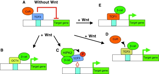
One possible explanation for these results comes from studies of OCT4, a key regulator of the pluripotency network (Babaie et al., 2007; Hanna et al., 2010; Jaenisch and Young, 2008; Loh et al., 2008; Niwa et al., 2000) (Fig. 4). β-catenin can form a complex with OCT4 to lead to the activation of OCT4-dependent reporters (Kelly et al., 2011; Takao et al., 2007) (Fig. 4B). Consistent with these observations, OCT4 has been proposed to promote the commitment of mouse ES cells to mesendoderm in response to Wnt signaling (Thomson et al., 2011). By contrast, a recent study found that β-catenin occupancy of OCT4-dependent promoters requires TCF proteins, implying that the effect of β-catenin on OCT4 targets is unlikely to be mediated by the direct interaction of β-catenin with OCT4 (Yi et al., 2011). The binding of OCT4 to β-catenin has been previously reported to antagonize Wnt pathway activity in axis formation (Abu-Remaileh et al., 2010; Cao et al., 2007), whereas its presumed stimulatory role in Wnt-dependent ES cell self-renewal awaits more direct experimental support.
Fig. 4.
Models for how Wnt signaling maintains pluripotency in ES cells. (A) In the absence of Wnt signaling, β-catenin (βcat) is degraded, and TCF3 in complex with transcriptional co-repressors (CoR) constitutively represses Wnt target genes. (B-E) Upon Wnt pathway activation, several alternative models leading to pluripotency are possible: (B) stabilized β-catenin associates with OCT4 to activate OCT4-dependent transcription; (C) HIPK2 is activated by Wnt signaling, associates with β-catenin and phosphorylates TCF3; this phosphorylation results in the removal of TCF3 from target promoters, leading to transcriptional derepression; (D) stabilized β-catenin associates with TCF3, causing the removal of the co-repressors resulting in target derepression (this model predicts that TCF3 is still bound to the promoter but no longer represses its gene targets); and (E) the TCF switch model, in which TCF3 repressor is replaced by TCF1 activator, leading to target activation and pluripotency. OCT4, octamer-binding transcription factor 4.
Inactivation of TCF3-mediated repression by Wnt signaling
Another explanation for how β-catenin might maintain ES cell pluripotency has recently emerged from studies into the role of TCF3 in Xenopus embryos (Hikasa et al., 2010). TCF3 is abundantly expressed during early vertebrate development and in ES cells (Hikasa et al., 2010; Kelly et al., 2011; Kim et al., 2000; Merrill et al., 2004; Pereira et al., 2006; Roel et al., 2002). In ES cells, TCF3 was found to occupy the same promoters as those occupied by the key pluripotency factors OCT4 and NANOG, and is likely to be responsible for much of the repression of ES cell-specific transcription (Cole et al., 2008; Tam et al., 2008; Yi et al., 2008). Loss-of-function studies in zebrafish, mouse and Xenopus embryos indicate that TCF3 is a transcriptional repressor (Hikasa et al., 2010; Houston et al., 2002; Kim et al., 2000; Merrill et al., 2004). Zebrafish headless embryos, which have a non-functional tcf3 (tcf7l1a) gene, have been rescued by the expression of a Tcf3-Engrailed fusion protein (Kim et al., 2000). Since the rescuing construct contains the Engrailed domain that functions to repress transcription, this result strongly suggests that Tcf3 is a transcriptional repressor. This conclusion contrasts with earlier observations, in which target genes were activated when TCF3 was complexed with β-catenin (Cole et al., 2008; Molenaar et al., 1996; van de Wetering et al., 1997). Tcf3–/– mouse ES cells show increased NANOG expression and do not undergo efficient differentiation in standard conditions, consistent with the view that TCF3 is a negative regulator of the ES cell pluripotency network (Cole et al., 2008; Pereira et al., 2006; Tam et al., 2008; Yi et al., 2008). In fact, TCF3 has been identified as an inhibitor of pluripotency in unbiased forward genetic screens in mouse ES cells (Guo et al., 2011; Schaniel et al., 2009) and during cell reprogramming (Han et al., 2010; Lluis et al., 2011). The majority of the genes repressed by TCF3 differ from those activated in response to Wnt proteins, indicating that TCF3 might have Wnt-independent functions (Cole et al., 2008; Merrill et al., 2004; Nguyen et al., 2006; Yi et al., 2011). Conversely, in Xenopus embryos, some Wnt/β-catenin targets, including the Cdx, Vent/Vox and Meis genes, are negatively controlled by TCF3 but are activated in response to Wnt proteins (Hikasa et al., 2010). The opposite regulation of ES cell self-renewal by TCF3 and Wnt signaling agonists has given rise to a hypothesis that the Wnt pathway acts to eliminate TCF3-mediated repression.
This hypothesis has been supported by the discovery of a specific, novel mechanism for Wnt signaling (Hikasa et al., 2010; Hikasa and Sokol, 2011) (Fig. 4C). TCF3 becomes rapidly phosphorylated in Xenopus ectoderm and in human embryonic kidney (HEK) 293T cells stimulated by WNT3A (Hikasa et al., 2010). Homeodomain-interacting protein kinase 2 (HIPK2) was identified in this study as being both necessary and sufficient for this phosphorylation. Additionally, β-catenin was found to serve as a scaffold that promotes TCF3 phosphorylation by attracting HIPK2 to TCF3 (Hikasa et al., 2010). This novel role of β-catenin is distinct from its commonly accepted function as a transcriptional coactivator. This phosphorylation of TCF3 occurred in response to the overexpression of LRP6 and upon inhibition of GSK3 and resulted in the removal of TCF3 from target promoters, including those of Vent2, Cdx4 and Siamois (Hikasa and Sokol, 2011; Hikasa et al., 2010) (Fig. 4C). Since the inactivation of TCF3 by phosphorylation occurs both in Xenopus and in mammalian cells (Hikasa et al., 2010; Hikasa and Sokol, 2011; Sokol, 2011), the phosphorylation model provides a potential molecular mechanism for the effects of GSK3 inhibition and loss of the Tcf3 gene on ES cell pluripotency and self-renewal (Kelly et al., 2011; Wray et al., 2011; Yi et al., 2011).
Consistent with this model, TCF3 depletion phenocopies the effect of GSK3 inhibitors on ES cell self-renewal (Wray et al., 2011; Yi et al., 2011). Similarly, the knockdown of Tcf3 by a short hairpin (sh) RNA rescued the clonogenic ability of ES cells, which was impaired by the loss of Ctnnb1 (Wray et al., 2011). Moreover, overexpression of either a stabilized or a C-terminally truncated β-catenin in mouse ES cells promoted their pluripotency and inhibited neuronal differentiation (Kelly et al., 2011). These findings suggest that this truncated form of β-catenin, although unable to activate a standard TCF reporter, acts by stimulating specific pluripotency target genes. Indeed, the truncated β-catenin rescued the expression of some TCF3 target genes in response to GSK3 inhibition (Wray et al., 2011). The TCF3 phosphorylation model predicts that the truncated β-catenin will serve as a scaffold for TCF3 phosphorylation, but this prediction remains to be tested.
By contrast, the effects of β-catenin on mouse ES cell pluripotency have not been abrogated by different dominant interfering TCF constructs, which was interpreted to suggest a TCF-independent mechanism for the self-renewal of these cells (Kelly et al., 2011). These TCF constructs retain the DNA-binding domain, but not the β-catenin-binding domain, and they can inhibit signaling by displacing a positively acting TCF from the target promoter. If ES cell pluripotency requires the physical removal of the TCF3 repressor from the target promoter, the binding of these constructs to target DNA will not prevent target activation. On the contrary, in the absence of a Wnt signal, the competition of these constructs with TCF3 might stimulate gene targets. This explanation leaves open the question of why the Wnt target genes Axin2, T and Cdx1, which should be repressed by TCF3, are efficiently downregulated by dominant interfering TCF4 in mouse ES cells (Kelly et al., 2011). Clearly, the interplay between the positive and negative TCF proteins that are expressed in early development (see below) should be considered when interpreting these findings. Also, the long-term effect of β-catenin on ES cell self-renewal could be more resistant to target gene inhibition by the truncated forms of TCF4 or TCF1 than direct transcriptional assays (Kelly et al., 2011). Of note, none of the above-mentioned interfering TCF constructs is related to TCF3. A similar construct for TCF3 behaves as a constitutive repressor (Hikasa et al., 2010) and would be expected to block the consequences of GSK3 inhibition (Wray et al., 2011). Thus, detailed molecular comparisons of TCF/LEF family members are needed to elucidate the protein domains responsible for the functional differences between TCF3 and the other TCF proteins.
Despite its attractiveness, the phosphorylation model does not explain all of the recent observations reported in this field. The Wnt/HIPK2-dependent phosphorylation of TCF3 has been shown to activate Wnt target genes that are responsible for posterior development (Cdx4, Vent2 and Meis) (Hikasa et al., 2010). However, Wnt target genes and TCF reporters were only poorly activated in ES cells depleted of TCF3 and in those that overexpress truncated β-catenin (Kelly et al., 2011; Lyashenko et al., 2011; Wray et al., 2011). In one study, the rescuing effect of the truncated β-catenin correlated with its ability to restore normal cell adhesion, suggesting that the function of β-catenin in cell adhesion, rather than in signaling, is crucial for germ layer formation in ES cell aggregates (Lyashenko et al., 2011). An alternative explanation for these findings is that only a subset of Wnt targets is activated by TCF3 derepression. The identification of specific TCF3 targets that are involved in pluripotency should shed light on the mechanism of their activation.
Another potential explanation for the mechanism of TCF3 derepression in ES cells is that β-catenin competes with Groucho co-repressors for TCF3 binding (Cavallo et al., 1998; Daniels and Weis, 2005; Roose et al., 1998) (Fig. 4D). A related mechanism has been proposed to operate in zebrafish embryonic development, although it has not been directly linked to Wnt signaling (Ro and Dawid, 2011). According to this model, TCF3 itself remains bound to its transcriptional targets, whereas the phosphorylation model predicts that TCF3 should be removed from promoter DNA (Fig. 4C). These mechanisms can be discriminated in chromatin immunoprecipitation studies by assessing TCF3 occupancy of relevant target promoters in ES cells. The co-repressor displacement model postulates that TCF3 represses target genes in the absence of a Wnt signal, but activates them upon Wnt stimulation. This is supported in Drosophila and C. elegans embryos, in which a single TCF gene plays both positive and negative roles in transcription (Brunner et al., 1997; Cavallo et al., 1998; Phillips and Kimble, 2009). Nevertheless, existing genetic evidence indicates that TCF3 acts as a transcriptional repressor rather than as an activator (Hikasa et al., 2010; Houston et al., 2002; Kim et al., 2000; Merrill et al., 2004). As shown in Table 1 and discussed further below, vertebrates have multiple TCF genes, making it essential to distinguish the roles of individual TCFs in embryonic development and in ES cell self-renewal.
Diverse roles of TCFs in Wnt signaling and ES cell pluripotency
Studies of vertebrate embryonic development have revealed that different TCF family members have diverse signaling roles (Arce et al., 2006; Gradl et al., 2002; Liu et al., 2005; Roel et al., 2002; Standley et al., 2006). Whereas TCF3 appears to function exclusively as a transcriptional repressor, TCF1, TCF4 and LEF1 are able to activate transcription (Galceran et al., 1999; Gradl et al., 2002; Hikasa and Sokol, 2011; Kim et al., 2000; Liu et al., 2005; Standley et al., 2006; van de Wetering et al., 1996; van Genderen et al., 1994). Given the dynamic and diverse expression patterns of TCF homologs in the vertebrate embryo (Archbold et al., 2011; Molenaar et al., 1998; Roel et al., 2003), it comes as no surprise that both specialized and redundant functions have been identified for vertebrate TCFs in loss-of-function studies (Table 1) (Bonner et al., 2008; Galceran et al., 1999; Gregorieff et al., 2004; Liu et al., 2005; Nagayoshi et al., 2008; Nguyen et al., 2009; Standley et al., 2006).
The presence of distinct TCFs in different embryonic tissues suggests that TCF proteins might be key factors in determining context-specific responses to Wnt signaling (Hoppler and Kavanagh, 2007). An important issue that warrants investigation is whether individual TCF proteins regulate the same or different sets of target genes and how this is achieved mechanistically. A recent study offers a possible mechanism to explain the opposite action of TCF3 and TCF1 on Vent2 gene regulation in Xenopus embryos (Hikasa and Sokol, 2011). These experiments revealed that the decreased binding of HIPK2-phosphorylated TCF3 to the Vent2 promoter is accompanied by the enhanced association of TCF1 to the same binding site, leading to further upregulation of transcriptional activity (Hikasa and Sokol, 2011) (Fig. 4E). Since all four TCF family members are present in mouse ES cells (Huang and Qin, 2010; Kelly et al., 2011; Pereira et al., 2006), similar functional interactions between the TCF proteins that control ES cell pluripotency are likely to occur. Consistent with this possibility, the expression of a Tcf1 shRNA in mouse ES cells has been reported to inhibit many of the same genes that are upregulated in mouse Tcf3–/– ES cells, indicating that TCF1 contributes to transcriptional activation in this system (Yi et al., 2011). Moreover, the response of Tcf3–/– ES cells to WNT3A stimulation is suppressed by this Tcf1 shRNA (Yi et al., 2011), in agreement with the increased association of TCF1 with TCF3 target promoters in TCF3-depleted cells (Hikasa and Sokol, 2011). Thus, the same TCF `switch' mechanism is likely to operate in both Xenopus embryonic cells and mouse ES cells, but this possibility remains to be tested experimentally.
Conclusions
Recent studies have uncovered a new mode of Wnt signaling that is relevant to the ability of this pathway to maintain pluripotency in ES cells and to regulate cell lineage and body axis specification in vertebrate embryos. ES cell pluripotency and self-renewal are promoted when the pathway is upregulated at any level, by adding exogenous WNT3A ligand, inhibiting GSK3 activity, overexpressing β-catenin or depleting TCF3. The emerging picture reveals two key molecular events downstream of Wnt signaling that synergize in promoting ES cell pluripotency through TCF3 inactivation. One event is the stabilization of β-catenin, the other is the inactivation of TCF3. In Xenopus embryos and HEK293 cells, TCF3 is inactivated by HIPK2-mediated phosphorylation in a process that requires β-catenin as a molecular scaffold rather than as a transcriptional coactivator. This conserved mechanism leads to TCF3 removal from target promoters and to transcriptional derepression. Although this pathway is the easiest way to explain the recent data on the regulation of pluripotency by GSK3, β-catenin and TCF3, it remains to be proven to operate in ES cells. Until a specific mechanism is demonstrated, it remains formally possible that TCF3 functions to control ES cell pluripotency in a Wnt-independent manner. Nevertheless, based on the available data, TCF3 phosphorylation appears to be responsible for both vertebrate AP axis specification and the maintenance of ES cell pluripotency, and might represent a new drug target for cell-based disease therapies.
Many of the existing controversies concerning the Wnt pathway and the maintenance of ES cell pluripotency predominantly arise from our having an incomplete understanding of the context- and stage-dependent functions of the TCF protein family members. Another gap in our knowledge concerns the alternative mechanisms of TCF3-dependent transcriptional derepression. This can be addressed by examining the role of transcriptional co-repressors in the signaling function of TCF proteins and assessing TCF occupancy of the relevant promoters during Wnt signaling. Yet another important question concerns the different molecular mechanisms that are employed to control distinct Wnt target genes. Only a subset of WNT3A and TCF3 gene targets appears to be shared, indicating that TCF3 has both Wnt-dependent and Wnt-independent functions (Cole et al., 2008; Yi et al., 2011). Thus, an important future goal is to identify specific target genes that are responsible for maintaining ES cell pluripotency. Although the current literature indicates that these targets are stimulated by Wnt proteins and are repressed by TCF3, it is also possible that Wnt ligands and downstream signaling components affect pluripotency via distinct gene targets. Further studies are needed to address these questions and to gain a better control over the balance between the self-renewal and the differentiation of ES cells.
Acknowledgments
I thank Julian Gingold, Christoph Schaniel, Jianlong Wang, Stephen Dalton and the three anonymous reviewers for critical comments on the manuscript, and the Brad Merrill and Austin Smith groups for sharing data prior to publication. I apologize to those authors whose work has not been cited owing to space constraints.
Footnotes
Funding
The work in the S.Y.S. laboratory is supported by NIH grants. Deposited in PMC for release after 12 months.
Competing interests statement
The authors declare no competing financial interests.
References
- Abu-Remaileh M., Gerson A., Farago M., Nathan G., Alkalay I., Zins Rousso S., Gur M., Fainsod A., Bergman Y. (2010). Oct-3/4 regulates stem cell identity and cell fate decisions by modulating Wnt/beta-catenin signalling. EMBO J. 29, 3236-3248 [DOI] [PMC free article] [PubMed] [Google Scholar]
- Angers S., Moon R. T. (2009). Proximal events in Wnt signal transduction. Nat. Rev. Mol. Cell Biol. 10, 468-477 [DOI] [PubMed] [Google Scholar]
- Anton R., Kestler H. A., Kuhl M. (2007). Beta-catenin signaling contributes to stemness and regulates early differentiation in murine embryonic stem cells. FEBS Lett. 581, 5247-5254 [DOI] [PubMed] [Google Scholar]
- Arce L., Yokoyama N. N., Waterman M. L. (2006). Diversity of LEF/TCF action in development and disease. Oncogene 25, 7492-7504 [DOI] [PubMed] [Google Scholar]
- Archbold H. C., Yang Y. X., Chen L., Cadigan K. M. (2011). How do they do Wnt they do?: regulation of transcription by the Wnt/beta-catenin pathway. Acta Physiol. doi: 10.1111/j.1748-1716.2011.02293.x [DOI] [PubMed]
- Arnold S. J., Robertson E. J. (2009). Making a commitment: cell lineage allocation and axis patterning in the early mouse embryo. Nat. Rev. Mol. Cell Biol. 10, 91-103 [DOI] [PubMed] [Google Scholar]
- Aubert J., Dunstan H., Chambers I., Smith A. (2002). Functional gene screening in embryonic stem cells implicates Wnt antagonism in neural differentiation. Nat. Biotechnol. 20, 1240-1245 [DOI] [PubMed] [Google Scholar]
- Babaie Y., Herwig R., Greber B., Brink T. C., Wruck W., Groth D., Lehrach H., Burdon T., Adjaye J. (2007). Analysis of Oct4-dependent transcriptional networks regulating self-renewal and pluripotency in human embryonic stem cells. Stem Cells 25, 500-510 [DOI] [PubMed] [Google Scholar]
- Bakre M. M., Hoi A., Mong J. C., Koh Y. Y., Wong K. Y., Stanton L. W. (2007). Generation of multipotential mesendodermal progenitors from mouse embryonic stem cells via sustained Wnt pathway activation. J. Biol. Chem. 282, 31703-31712 [DOI] [PubMed] [Google Scholar]
- Bonner J., Gribble S. L., Veien E. S., Nikolaus O. B., Weidinger G., Dorsky R. I. (2008). Proliferation and patterning are mediated independently in the dorsal spinal cord downstream of canonical Wnt signaling. Dev. Biol. 313, 398-407 [DOI] [PMC free article] [PubMed] [Google Scholar]
- Boyer L. A., Lee T. I., Cole M. F., Johnstone S. E., Levine S. S., Zucker J. P., Guenther M. G., Kumar R. M., Murray H. L., Jenner R. G., et al. (2005). Core transcriptional regulatory circuitry in human embryonic stem cells. Cell 122, 947-956 [DOI] [PMC free article] [PubMed] [Google Scholar]
- Brockes J. P., Kumar A. (2008). Comparative aspects of animal regeneration. Annu. Rev. Cell Dev. Biol. 24, 525-549 [DOI] [PubMed] [Google Scholar]
- Brunner E., Peter O., Schweizer L., Basler K. (1997). pangolin encodes a Lef-1 homologue that acts downstream of Armadillo to transduce the Wingless signal in Drosophila. Nature 385, 829-833 [DOI] [PubMed] [Google Scholar]
- Cao Y., Siegel D., Donow C., Knochel S., Yuan L., Knochel W. (2007). POU-V factors antagonize maternal VegT activity and beta-Catenin signaling in Xenopus embryos. EMBO J. 26, 2942-2954 [DOI] [PMC free article] [PubMed] [Google Scholar]
- Cavallo R. A., Cox R. T., Moline M. M., Roose J., Polevoy G. A., Clevers H., Peifer M., Bejsovec A. (1998). Drosophila Tcf and Groucho interact to repress Wingless signalling activity. Nature 395, 604-608 [DOI] [PubMed] [Google Scholar]
- Chambers I., Colby D., Robertson M., Nichols J., Lee S., Tweedie S., Smith A. (2003). Functional expression cloning of Nanog, a pluripotency sustaining factor in embryonic stem cells. Cell 113, 643-655 [DOI] [PubMed] [Google Scholar]
- Clevers H. (2006). Wnt/beta-catenin signaling in development and disease. Cell 127, 469-480 [DOI] [PubMed] [Google Scholar]
- Cole M. F., Johnstone S. E., Newman J. J., Kagey M. H., Young R. A. (2008). Tcf3 is an integral component of the core regulatory circuitry of embryonic stem cells. Genes Dev. 22, 746-755 [DOI] [PMC free article] [PubMed] [Google Scholar]
- Conlon F. L., Lyons K. M., Takaesu N., Barth K. S., Kispert A., Herrmann B., Robertson E. J. (1994). A primary requirement for nodal in the formation and maintenance of the primitive streak in the mouse. Development 120, 1919-1928 [DOI] [PubMed] [Google Scholar]
- Daniels D. L., Weis W. I. (2005). Beta-catenin directly displaces Groucho/TLE repressors from Tcf/Lef in Wnt-mediated transcription activation. Nat. Struct. Mol. Biol. 12, 364-371 [DOI] [PubMed] [Google Scholar]
- Doble B. W., Woodgett J. R. (2003). GSK-3: tricks of the trade for a multi-tasking kinase. J. Cell Sci. 116, 1175-1186 [DOI] [PMC free article] [PubMed] [Google Scholar]
- Doble B. W., Patel S., Wood G. A., Kockeritz L. K., Woodgett J. R. (2007). Functional redundancy of GSK-3alpha and GSK-3beta in Wnt/beta-catenin signaling shown by using an allelic series of embryonic stem cell lines. Dev. Cell 12, 957-971 [DOI] [PMC free article] [PubMed] [Google Scholar]
- Dravid G., Ye Z., Hammond H., Chen G., Pyle A., Donovan P., Yu X., Cheng L. (2005). Defining the role of Wnt/beta-catenin signaling in the survival, proliferation, and self-renewal of human embryonic stem cells. Stem Cells 23, 1489-1501 [DOI] [PubMed] [Google Scholar]
- Evans M. J., Kaufman M. H. (1981). Establishment in culture of pluripotential cells from mouse embryos. Nature 292, 154-156 [DOI] [PubMed] [Google Scholar]
- Foley A. C., Mercola M. (2005). Heart induction by Wnt antagonists depends on the homeodomain transcription factor Hex. Genes Dev. 19, 387-396 [DOI] [PMC free article] [PubMed] [Google Scholar]
- Gadue P., Huber T. L., Paddison P. J., Keller G. M. (2006). Wnt and TGF-beta signaling are required for the induction of an in vitro model of primitive streak formation using embryonic stem cells. Proc. Natl. Acad. Sci. USA 103, 16806-16811 [DOI] [PMC free article] [PubMed] [Google Scholar]
- Galceran J., Farinas I., Depew M. J., Clevers H., Grosschedl R. (1999). Wnt3a–/–-like phenotype and limb deficiency in Lef1–/–Tcf1–/– mice. Genes Dev. 13, 709-717 [DOI] [PMC free article] [PubMed] [Google Scholar]
- Gardner R. L., Meredith M. R., Altman D. G. (1992). Is the anterior-posterior axis of the fetus specified before implantation in the mouse? J. Exp. Zool. 264, 437-443 [DOI] [PubMed] [Google Scholar]
- Gradl D., Konig A., Wedlich D. (2002). Functional diversity of Xenopus lymphoid enhancer factor/T-cell factor transcription factors relies on combinations of activating and repressing elements. J. Biol. Chem. 277, 14159-14171 [DOI] [PubMed] [Google Scholar]
- Gregorieff A., Grosschedl R., Clevers H. (2004). Hindgut defects and transformation of the gastro-intestinal tract in Tcf4–/–/Tcf1–/– embryos. EMBO J. 23, 1825-1833 [DOI] [PMC free article] [PubMed] [Google Scholar]
- Gribble S. L., Kim H. S., Bonner J., Wang X., Dorsky R. I. (2009). Tcf3 inhibits spinal cord neurogenesis by regulating sox4a expression. Development 136, 781-789 [DOI] [PMC free article] [PubMed] [Google Scholar]
- Grigoryan T., Wend P., Klaus A., Birchmeier W. (2008). Deciphering the function of canonical Wnt signals in development and disease: conditional loss- and gain-of-function mutations of beta-catenin in mice. Genes Dev. 22, 2308-2341 [DOI] [PMC free article] [PubMed] [Google Scholar]
- Guo G., Huang Y., Humphreys P., Wang X., Smith A. (2011). A PiggyBac-based recessive screening method to identify pluripotency regulators. PLoS ONE 6, e18189 [DOI] [PMC free article] [PubMed] [Google Scholar]
- Haegel H., Larue L., Ohsugi M., Fedorov L., Herrenknecht K., Kemler R. (1995). Lack of beta-catenin affects mouse development at gastrulation. Development 121, 3529-3537 [DOI] [PubMed] [Google Scholar]
- Haegele L., Ingold B., Naumann H., Tabatabai G., Ledermann B., Brandner S. (2003). Wnt signalling inhibits neural differentiation of embryonic stem cells by controlling bone morphogenetic protein expression. Mol. Cell. Neurosci. 24, 696-708 [DOI] [PubMed] [Google Scholar]
- Han J., Yuan P., Yang H., Zhang J., Soh B. S., Li P., Lim S. L., Cao S., Tay J., Orlov Y. L., et al. (2010). Tbx3 improves the germ-line competency of induced pluripotent stem cells. Nature 463, 1096-1100 [DOI] [PMC free article] [PubMed] [Google Scholar]
- Hanna J. H., Saha K., Jaenisch R. (2010). Pluripotency and cellular reprogramming: facts, hypotheses, unresolved issues. Cell 143, 508-525 [DOI] [PMC free article] [PubMed] [Google Scholar]
- Hao J., Li T. G., Qi X., Zhao D. F., Zhao G. Q. (2006). WNT/beta-catenin pathway up-regulates Stat3 and converges on LIF to prevent differentiation of mouse embryonic stem cells. Dev. Biol. 290, 81-91 [DOI] [PubMed] [Google Scholar]
- Harland R., Gerhart J. (1997). Formation and function of Spemann’s organizer. Annu. Rev. Cell Dev. Biol. 13, 611-667 [DOI] [PubMed] [Google Scholar]
- Heasman J., Crawford A., Goldstone K., Garner-Hamrick P., Gumbiner B., McCrea P., Kintner C., Noro C. Y., Wylie C. (1994). Overexpression of cadherins and underexpression of beta-catenin inhibit dorsal mesoderm induction in early Xenopus embryos. Cell 79, 791-803 [DOI] [PubMed] [Google Scholar]
- Heeg-Truesdell E., LaBonne C. (2006). Neural induction in Xenopus requires inhibition of Wnt-beta-catenin signaling. Dev. Biol. 298, 71-86 [DOI] [PubMed] [Google Scholar]
- Hikasa H., Sokol S. Y. (2011). Phosphorylation of TCF proteins by homeodomain-interacting protein kinase 2. J. Biol. Chem. 286, 12093-12100 [DOI] [PMC free article] [PubMed] [Google Scholar]
- Hikasa H., Ezan J., Itoh K., Li X., Klymkowsky M. W., Sokol S. Y. (2010). Regulation of TCF3 by Wnt-dependent phosphorylation during vertebrate axis specification. Dev. Cell 19, 521-532 [DOI] [PMC free article] [PubMed] [Google Scholar]
- Hoppler S., Moon R. T. (1998). BMP-2/-4 and Wnt-8 cooperatively pattern the Xenopus mesoderm. Mech. Dev. 71, 119-129 [DOI] [PubMed] [Google Scholar]
- Hoppler S., Kavanagh C. L. (2007). Wnt signalling: variety at the core. J. Cell Sci. 120, 385-393 [DOI] [PubMed] [Google Scholar]
- Houston D. W., Kofron M., Resnik E., Langland R., Destree O., Wylie C., Heasman J. (2002). Repression of organizer genes in dorsal and ventral Xenopus cells mediated by maternal XTcf3. Development 129, 4015-4025 [DOI] [PubMed] [Google Scholar]
- Hsu S. C., Galceran J., Grosschedl R. (1998). Modulation of transcriptional regulation by LEF-1 in response to Wnt-1 signaling and association with beta-catenin. Mol. Cell. Biol. 18, 4807-4818 [DOI] [PMC free article] [PubMed] [Google Scholar]
- Huang C., Qin D. (2010). Role of Lef1 in sustaining self-renewal in mouse embryonic stem cells. J. Genet. Genomics 37, 441-449 [DOI] [PubMed] [Google Scholar]
- Huelsken J., Vogel R., Brinkmann V., Erdmann B., Birchmeier C., Birchmeier W. (2000). Requirement for beta-catenin in anterior-posterior axis formation in mice. J. Cell Biol. 148, 567-578 [DOI] [PMC free article] [PubMed] [Google Scholar]
- Itoh K., Sokol S. Y. (1999). Axis determination by inhibition of Wnt signaling in Xenopus. Genes Dev. 13, 2328-2336 [DOI] [PMC free article] [PubMed] [Google Scholar]
- Jaenisch R., Young R. (2008). Stem cells, the molecular circuitry of pluripotency and nuclear reprogramming. Cell 132, 567-582 [DOI] [PMC free article] [PubMed] [Google Scholar]
- Kelly K. F., Ng D. Y., Jayakumaran G., Wood G. A., Koide H., Doble B. W. (2011). beta-catenin enhances Oct-4 activity and reinforces pluripotency through a TCF-independent mechanism. Cell Stem Cell 8, 214-227 [DOI] [PMC free article] [PubMed] [Google Scholar]
- Kelly O. G., Pinson K. I., Skarnes W. C. (2004). The Wnt co-receptors Lrp5 and Lrp6 are essential for gastrulation in mice. Development 131, 2803-2815 [DOI] [PubMed] [Google Scholar]
- Kielman M. F., Rindapaa M., Gaspar C., van Poppel N., Breukel C., van Leeuwen S., Taketo M. M., Roberts S., Smits R., Fodde R. (2002). Apc modulates embryonic stem-cell differentiation by controlling the dosage of beta-catenin signaling. Nat. Genet. 32, 594-605 [DOI] [PubMed] [Google Scholar]
- Kim C. H., Oda T., Itoh M., Jiang D., Artinger K. B., Chandrasekharappa S. C., Driever W., Chitnis A. B. (2000). Repressor activity of Headless/Tcf3 is essential for vertebrate head formation. Nature 407, 913-916 [DOI] [PMC free article] [PubMed] [Google Scholar]
- Kolligs F. T., Hu G., Dang C. V., Fearon E. R. (1999). Neoplastic transformation of RK3E by mutant beta-catenin requires deregulation of Tcf/Lef transcription but not activation of c-myc expression. Mol. Cell. Biol. 19, 5696-5706 [DOI] [PMC free article] [PubMed] [Google Scholar]
- Korinek V., Barker N., Moerer P., van Donselaar E., Huls G., Peters P. J., Clevers H. (1998). Depletion of epithelial stem-cell compartments in the small intestine of mice lacking Tcf-4. Nat. Genet. 19, 379-383 [DOI] [PubMed] [Google Scholar]
- Kragl M., Knapp D., Nacu E., Khattak S., Maden M., Epperlein H. H., Tanaka E. M. (2009). Cells keep a memory of their tissue origin during axolotl limb regeneration. Nature 460, 60-65 [DOI] [PubMed] [Google Scholar]
- Kunz M., Herrmann M., Wedlich D., Gradl D. (2004). Autoregulation of canonical Wnt signaling controls midbrain development. Dev. Biol. 273, 390-401 [DOI] [PubMed] [Google Scholar]
- Lindsley R. C., Gill J. G., Kyba M., Murphy T. L., Murphy K. M. (2006). Canonical Wnt signaling is required for development of embryonic stem cell-derived mesoderm. Development 133, 3787-3796 [DOI] [PubMed] [Google Scholar]
- Liu F., van den Broek O., Destree O., Hoppler S. (2005). Distinct roles for Xenopus Tcf/Lef genes in mediating specific responses to Wnt/beta-catenin signalling in mesoderm development. Development 132, 5375-5385 [DOI] [PubMed] [Google Scholar]
- Liu P., Wakamiya M., Shea M. J., Albrecht U., Behringer R. R., Bradley A. (1999). Requirement for Wnt3 in vertebrate axis formation. Nat. Genet. 22, 361-365 [DOI] [PubMed] [Google Scholar]
- Lluis F., Pedone E., Pepe S., Cosma M. P. (2008). Periodic activation of Wnt/beta-catenin signaling enhances somatic cell reprogramming mediated by cell fusion. Cell Stem Cell 3, 493-507 [DOI] [PubMed] [Google Scholar]
- Lluis F., Ombrato L., Pedone E., Pepe S., Merrill B. J., Cosma M. P. (2011). T-cell factor 3 (Tcf3) deletion increases somatic cell reprogramming by inducing epigenome modifications. Proc. Natl. Acad. Sci. USA 108, 11912-11917 [DOI] [PMC free article] [PubMed] [Google Scholar]
- Loh Y. H., Wu Q., Chew J. L., Vega V. B., Zhang W., Chen X., Bourque G., George J., Leong B., Liu J., et al. (2006). The Oct4 and Nanog transcription network regulates pluripotency in mouse embryonic stem cells. Nat. Genet. 38, 431-440 [DOI] [PubMed] [Google Scholar]
- Loh Y. H., Ng J. H., Ng H. H. (2008). Molecular framework underlying pluripotency. Cell Cycle 7, 885-891 [DOI] [PubMed] [Google Scholar]
- Lyashenko N., Winter M., Migliorini D., Biechele T., Moon R. T., Hartmann C. (2011). Differential requirement for the dual functions of beta-catenin in embryonic stem cell self-renewal and germ layer formation. Nat. Cell Biol. 13, 753-761 [DOI] [PMC free article] [PubMed] [Google Scholar]
- MacDonald B. T., Tamai K., He X. (2009). Wnt/beta-catenin signaling: components, mechanisms, and diseases. Dev. Cell 17, 9-26 [DOI] [PMC free article] [PubMed] [Google Scholar]
- Marson A., Foreman R., Chevalier B., Bilodeau S., Kahn M., Young R. A., Jaenisch R. (2008). Wnt signaling promotes reprogramming of somatic cells to pluripotency. Cell Stem Cell 3, 132-135 [DOI] [PMC free article] [PubMed] [Google Scholar]
- Martin G. R. (1981). Isolation of a pluripotent cell line from early mouse embryos cultured in medium conditioned by teratocarcinoma stem cells. Proc. Natl. Acad. Sci. USA 78, 7634-7638 [DOI] [PMC free article] [PubMed] [Google Scholar]
- McGrew L. L., Hoppler S., Moon R. T. (1997). Wnt and FGF pathways cooperatively pattern anteroposterior neural ectoderm in Xenopus. Mech. Dev. 69, 105-114 [DOI] [PubMed] [Google Scholar]
- Merrill B. J., Pasolli H. A., Polak L., Rendl M., Garcia-Garcia M. J., Anderson K. V., Fuchs E. (2004). Tcf3: a transcriptional regulator of axis induction in the early embryo. Development 131, 263-274 [DOI] [PubMed] [Google Scholar]
- Mitsui K., Tokuzawa Y., Itoh H., Segawa K., Murakami M., Takahashi K., Maruyama M., Maeda M., Yamanaka S. (2003). The homeoprotein Nanog is required for maintenance of pluripotency in mouse epiblast and ES cells. Cell 113, 631-642 [DOI] [PubMed] [Google Scholar]
- Miyabayashi T., Teo J. L., Yamamoto M., McMillan M., Nguyen C., Kahn M. (2007). Wnt/beta-catenin/CBP signaling maintains long-term murine embryonic stem cell pluripotency. Proc. Natl. Acad. Sci. USA 104, 5668-5673 [DOI] [PMC free article] [PubMed] [Google Scholar]
- Molenaar M., van de Wetering M., Oosterwegel M., Peterson-Maduro J., Godsave S., Korinek V., Roose J., Destree O., Clevers H. (1996). XTcf-3 transcription factor mediates beta-catenin-induced axis formation in Xenopus embryos. Cell 86, 391-399 [DOI] [PubMed] [Google Scholar]
- Molenaar M., Roose J., Peterson J., Venanzi S., Clevers H., Destree O. (1998). Differential expression of the HMG box transcription factors XTcf-3 and XLef-1 during early Xenopus development. Mech. Dev. 75, 151-154 [DOI] [PubMed] [Google Scholar]
- Mukhopadhyay M., Shtrom S., Rodriguez-Esteban C., Chen L., Tsukui T., Gomer L., Dorward D. W., Glinka A., Grinberg A., Huang S. P., et al. (2001). Dickkopf1 is required for embryonic head induction and limb morphogenesis in the mouse. Dev. Cell 1, 423-434 [DOI] [PubMed] [Google Scholar]
- Muncan V., Faro A., Haramis A. P., Hurlstone A. F., Wienholds E., van Es J., Korving J., Begthel H., Zivkovic D., Clevers H. (2007). T-cell factor 4 (Tcf7l2) maintains proliferative compartments in zebrafish intestine. EMBO Rep. 8, 966-973 [DOI] [PMC free article] [PubMed] [Google Scholar]
- Nagayoshi S., Hayashi E., Abe G., Osato N., Asakawa K., Urasaki A., Horikawa K., Ikeo K., Takeda H., Kawakami K. (2008). Insertional mutagenesis by the Tol2 transposon-mediated enhancer trap approach generated mutations in two developmental genes: tcf7 and synembryn-like. Development 135, 159-169 [DOI] [PubMed] [Google Scholar]
- Naito A. T., Shiojima I., Akazawa H., Hidaka K., Morisaki T., Kikuchi A., Komuro I. (2006). Developmental stage-specific biphasic roles of Wnt/beta-catenin signaling in cardiomyogenesis and hematopoiesis. Proc. Natl. Acad. Sci. USA 103, 19812-19817 [DOI] [PMC free article] [PubMed] [Google Scholar]
- Nguyen H., Rendl M., Fuchs E. (2006). Tcf3 governs stem cell features and represses cell fate determination in skin. Cell 127, 171-183 [DOI] [PubMed] [Google Scholar]
- Nguyen H., Merrill B. J., Polak L., Nikolova M., Rendl M., Shaver T. M., Pasolli H. A., Fuchs E. (2009). Tcf3 and Tcf4 are essential for long-term homeostasis of skin epithelia. Nat. Genet. 41, 1068-1075 [DOI] [PMC free article] [PubMed] [Google Scholar]
- Nichols J., Smith A. (2011). The origin and identity of embryonic stem cells. Development 138, 3-8 [DOI] [PubMed] [Google Scholar]
- Niwa H., Miyazaki J., Smith A. G. (2000). Quantitative expression of Oct-3/4 defines differentiation, dedifferentiation or self-renewal of ES cells. Nat. Genet. 24, 372-376 [DOI] [PubMed] [Google Scholar]
- Nusse R., Fuerer C., Ching W., Harnish K., Logan C., Zeng A., ten Berge D., Kalani Y. (2008). Wnt signaling and stem cell control. Cold Spring Harb. Symp. Quant. Biol. 73, 59-66 [DOI] [PubMed] [Google Scholar]
- Ogawa K., Nishinakamura R., Iwamatsu Y., Shimosato D., Niwa H. (2006). Synergistic action of Wnt and LIF in maintaining pluripotency of mouse ES cells. Biochem. Biophys. Res. Commun. 343, 159-166 [DOI] [PubMed] [Google Scholar]
- Pandur P., Lasche M., Eisenberg L. M., Kuhl M. (2002). Wnt-11 activation of a non-canonical Wnt signalling pathway is required for cardiogenesis. Nature 418, 636-641 [DOI] [PubMed] [Google Scholar]
- Pereira L., Yi F., Merrill B. J. (2006). Repression of Nanog gene transcription by Tcf3 limits embryonic stem cell self-renewal. Mol. Cell. Biol. 26, 7479-7491 [DOI] [PMC free article] [PubMed] [Google Scholar]
- Petersen C. P., Reddien P. W. (2009). Wnt signaling and the polarity of the primary body axis. Cell 139, 1056-1068 [DOI] [PubMed] [Google Scholar]
- Phillips B. T., Kimble J. (2009). A new look at TCF and beta-catenin through the lens of a divergent C. elegans Wnt pathway. Dev. Cell 17, 27-34 [DOI] [PMC free article] [PubMed] [Google Scholar]
- Reya T., O’Riordan M., Okamura R., Devaney E., Willert K., Nusse R., Grosschedl R. (2000). Wnt signaling regulates B lymphocyte proliferation through a LEF-1 dependent mechanism. Immunity 13, 15-24 [DOI] [PubMed] [Google Scholar]
- Ro H., Dawid I. B. (2011). Modulation of Tcf3 repressor complex composition regulates cdx4 expression in zebrafish. EMBO J. 30, 2894-2907 [DOI] [PMC free article] [PubMed] [Google Scholar]
- Roel G., Hamilton F. S., Gent Y., Bain A. A., Destree O., Hoppler S. (2002). Lef-1 and Tcf-3 transcription factors mediate tissue-specific Wnt signaling during Xenopus development. Curr. Biol. 12, 1941-1945 [DOI] [PubMed] [Google Scholar]
- Roel G., van den Broek O., Spieker N., Peterson-Maduro J., Destree O. (2003). Tcf-1 expression during Xenopus development. Gene Expr. Patterns 3, 123-126 [DOI] [PubMed] [Google Scholar]
- Roose J., Molenaar M., Peterson J., Hurenkamp J., Brantjes H., Moerer P., van de Wetering M., Destree O., Clevers H. (1998). The Xenopus Wnt effector XTcf-3 interacts with Groucho-related transcriptional repressors. Nature 395, 608-612 [DOI] [PubMed] [Google Scholar]
- Roose J., Huls G., van Beest M., Moerer P., van der Horn K., Goldschmeding R., Logtenberg T., Clevers H. (1999). Synergy between tumor suppressor APC and the beta-catenin-Tcf4 target Tcf1. Science 285, 1923-1926 [DOI] [PubMed] [Google Scholar]
- Rosner M. H., Vigano M. A., Ozato K., Timmons P. M., Poirier F., Rigby P. W., Staudt L. M. (1990). A POU-domain transcription factor in early stem cells and germ cells of the mammalian embryo. Nature 345, 686-692 [DOI] [PubMed] [Google Scholar]
- Rossant J., Tam P. P. (2009). Blastocyst lineage formation, early embryonic asymmetries and axis patterning in the mouse. Development 136, 701-713 [DOI] [PubMed] [Google Scholar]
- Sato N., Meijer L., Skaltsounis L., Greengard P., Brivanlou A. H. (2004). Maintenance of pluripotency in human and mouse embryonic stem cells through activation of Wnt signaling by a pharmacological GSK-3-specific inhibitor. Nat. Med. 10, 55-63 [DOI] [PubMed] [Google Scholar]
- Savage R., Phillips C. R. (1989). Signals from the dorsal blastopore lip region during gastrulation bias the ectoderm toward a nonepidermal pathway of differentiation in Xenopus laevis. Dev. Biol. 133, 157-168 [DOI] [PubMed] [Google Scholar]
- Schaniel C., Ang Y. S., Ratnakumar K., Cormier C., James T., Bernstein E., Lemischka I. R., Paddison P. J. (2009). Smarcc1/Baf155 couples self-renewal gene repression with changes in chromatin structure in mouse embryonic stem cells. Stem Cells 27, 2979-2991 [DOI] [PMC free article] [PubMed] [Google Scholar]
- Schier A. F., Talbot W. S. (2005). Molecular genetics of axis formation in zebrafish. Annu. Rev. Genet. 39, 561-613 [DOI] [PubMed] [Google Scholar]
- Singla D. K., Schneider D. J., LeWinter M. M., Sobel B. E. (2006). wnt3a but not wnt11 supports self-renewal of embryonic stem cells. Biochem. Biophys. Res. Commun. 345, 789-795 [DOI] [PubMed] [Google Scholar]
- Smith A. G., Heath J. K., Donaldson D. D., Wong G. G., Moreau J., Stahl M., Rogers D. (1988). Inhibition of pluripotential embryonic stem cell differentiation by purified polypeptides. Nature 336, 688-690 [DOI] [PubMed] [Google Scholar]
- Sokol S., Melton D. A. (1991). Pre-existent pattern in Xenopus animal pole cells revealed by induction with activin. Nature 351, 409-411 [DOI] [PubMed] [Google Scholar]
- Sokol S. Y. (1993). Mesoderm formation in Xenopus ectodermal explants overexpressing Xwnt8: evidence for a cooperating signal reaching the animal pole by gastrulation. Development 118, 1335-1342 [DOI] [PubMed] [Google Scholar]
- Sokol S. Y. (1999). Wnt signaling and dorso-ventral axis specification in vertebrates. Curr. Opin. Genet. Dev. 9, 405-410 [DOI] [PubMed] [Google Scholar]
- Sokol S. Y. (2011). Wnt signaling through T-cell factor phosphorylation. Cell Res. 21, 1002-1012 [DOI] [PMC free article] [PubMed] [Google Scholar]
- Sokol S. Y., Melton D. A. (1992). Interaction of Wnt and activin in dorsal mesoderm induction in Xenopus. Dev. Biol. 154, 348-355 [DOI] [PubMed] [Google Scholar]
- Standley H. J., Destree O., Kofron M., Wylie C., Heasman J. (2006). Maternal XTcf1 and XTcf4 have distinct roles in regulating Wnt target genes. Dev. Biol. 289, 318-328 [DOI] [PubMed] [Google Scholar]
- Takada S., Stark K. L., Shea M. J., Vassileva G., McMahon J. A., McMahon A. P. (1994). Wnt-3a regulates somite and tailbud formation in the mouse embryo. Genes Dev. 8, 174-189 [DOI] [PubMed] [Google Scholar]
- Takao Y., Yokota T., Koide H. (2007). Beta-catenin up-regulates Nanog expression through interaction with Oct-3/4 in embryonic stem cells. Biochem. Biophys. Res. Commun. 353, 699-705 [DOI] [PubMed] [Google Scholar]
- Tam W. L., Lim C. Y., Han J., Zhang J., Ang Y. S., Ng H. H., Yang H., Lim B. (2008). T-cell factor 3 regulates embryonic stem cell pluripotency and self-renewal by the transcriptional control of multiple lineage pathways. Stem Cells 26, 2019-2031 [DOI] [PMC free article] [PubMed] [Google Scholar]
- ten Berge D., Brugmann S. A., Helms J. A., Nusse R. (2008a). Wnt and FGF signals interact to coordinate growth with cell fate specification during limb development. Development 135, 3247-3257 [DOI] [PMC free article] [PubMed] [Google Scholar]
- ten Berge D., Koole W., Fuerer C., Fish M., Eroglu E., Nusse R. (2008b). Wnt signaling mediates self-organization and axis formation in embryoid bodies. Cell Stem Cell 3, 508-518 [DOI] [PMC free article] [PubMed] [Google Scholar]
- ten Berge D., Kurek D., Siu R., Nusse R. (2011). Wnt proteins control the murine embryonic to epiblast stem cell transition. Nat. Cell Biol. 13, 1070-1075 [DOI] [PMC free article] [PubMed] [Google Scholar]
- Thomson M., Liu S. J., Zou L.-N., Smith Z., Meissner A., Ramanathan S. (2011). Pluripotency factors in embryonic stem cells regulate differentiation into germ layers. Cell 145, 875-889 [DOI] [PMC free article] [PubMed] [Google Scholar]
- Tzahor E., Lassar A. B. (2001). Wnt signals from the neural tube block ectopic cardiogenesis. Genes Dev. 15, 255-260 [DOI] [PMC free article] [PubMed] [Google Scholar]
- Ueno S., Weidinger G., Osugi T., Kohn A. D., Golob J. L., Pabon L., Reinecke H., Moon R. T., Murry C. E. (2007). Biphasic role for Wnt/beta-catenin signaling in cardiac specification in zebrafish and embryonic stem cells. Proc. Natl. Acad. Sci. USA 104, 9685-9690 [DOI] [PMC free article] [PubMed] [Google Scholar]
- van Amerongen R., Nusse R. (2009). Towards an integrated view of Wnt signaling in development. Development 136, 3205-3214 [DOI] [PubMed] [Google Scholar]
- van de Wetering M., Castrop J., Korinek V., Clevers H. (1996). Extensive alternative splicing and dual promoter usage generate Tcf-1 protein isoforms with differential transcription control properties. Mol. Cell. Biol. 16, 745-752 [DOI] [PMC free article] [PubMed] [Google Scholar]
- van de Wetering M., Cavallo R., Dooijes D., van Beest M., van Es J., Loureiro J., Ypma A., Hursh D., Jones T., Bejsovec A., et al. (1997). Armadillo coactivates transcription driven by the product of the Drosophila segment polarity gene dTCF. Cell 88, 789-799 [DOI] [PubMed] [Google Scholar]
- van den Berg D. L., Snoek T., Mullin N. P., Yates A., Bezstarosti K., Demmers J., Chambers I., Poot R. A. (2010). An Oct4-centered protein interaction network in embryonic stem cells. Cell Stem Cell 6, 369-381 [DOI] [PMC free article] [PubMed] [Google Scholar]
- van Genderen C., Okamura R. M., Farinas I., Quo R. G., Parslow T. G., Bruhn L., Grosschedl R. (1994). Development of several organs that require inductive epithelial-mesenchymal interactions is impaired in LEF-1-deficient mice. Genes Dev. 8, 2691-2703 [DOI] [PubMed] [Google Scholar]
- Verbeek S., Izon D., Hofhuis F., Robanus-Maandag E., te Riele H., van de Wetering M., Oosterwegel M., Wilson A., MacDonald H. R., Clevers H. (1995). An HMG-box-containing T-cell factor required for thymocyte differentiation. Nature 374, 70-74 [DOI] [PubMed] [Google Scholar]
- Wallingford J. B., Fraser S. E., Harland R. M. (2002). Convergent extension: the molecular control of polarized cell movement during embryonic development. Dev. Cell 2, 695-706 [DOI] [PubMed] [Google Scholar]
- Wend P., Holland J. D., Ziebold U., Birchmeier W. (2010). Wnt signaling in stem and cancer stem cells. Semin. Cell Dev. Biol. 21, 855-863 [DOI] [PubMed] [Google Scholar]
- Williams R. L., Hilton D. J., Pease S., Willson T. A., Stewart C. L., Gearing D. P., Wagner E. F., Metcalf D., Nicola N. A., Gough N. M. (1988). Myeloid leukaemia inhibitory factor maintains the developmental potential of embryonic stem cells. Nature 336, 684-687 [DOI] [PubMed] [Google Scholar]
- Wray J., Kalkan T., Smith A. G. (2011). Inhibition of glycogen synthase kinase 3 alleviates Tcf3 repression of the pluripotency. Nat. Cell Biol. 13, 838-845 [DOI] [PMC free article] [PubMed] [Google Scholar]
- Wu D., Pan W. (2010). GSK3: a multifaceted kinase in Wnt signaling. Trends Biochem. Sci. 35, 161-168 [DOI] [PMC free article] [PubMed] [Google Scholar]
- Yi F., Pereira L., Merrill B. J. (2008). Tcf3 functions as a steady-state limiter of transcriptional programs of mouse embryonic stem cell self-renewal. Stem Cells 26, 1951-1960 [DOI] [PMC free article] [PubMed] [Google Scholar]
- Yi F., Pereira L., Merrill B. J. (2011). Opposing effect of Tcf3 and Tcf1 control Wnt stimulation of ESC self-renewal. Nat. Cell Biol. 316, 1050-1060 [DOI] [PMC free article] [PubMed] [Google Scholar]
- Ying Q. L., Wray J., Nichols J., Batlle-Morera L., Doble B., Woodgett J., Cohen P., Smith A. (2008). The ground state of embryonic stem cell self-renewal. Nature 453, 519-523 [DOI] [PMC free article] [PubMed] [Google Scholar]
- Yoshikawa Y., Fujimori T., McMahon A. P., Takada S. (1997). Evidence that absence of Wnt-3a signaling promotes neuralization instead of paraxial mesoderm development in the mouse. Dev. Biol. 183, 234-242 [DOI] [PubMed] [Google Scholar]
- Zeng L., Fagotto F., Zhang T., Hsu W., Vasicek T. J., Perry W. L., 3rd, Lee J. J., Tilghman S. M., Gumbiner B. M., Costantini F. (1997). The mouse Fused locus encodes Axin, an inhibitor of the Wnt signaling pathway that regulates embryonic axis formation. Cell 90, 181-192 [DOI] [PubMed] [Google Scholar]


