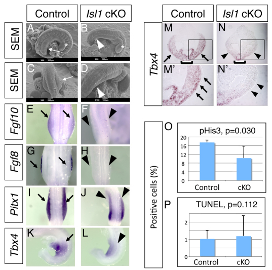
Fig. 3.
Islet1 is required for hindlimb initiation in the mouse embryo. (A-L) Scanning electron microscopy analysis (A-D) and whole-mount mRNA expression analysis (E-L) of Islet1 cKO embryos (B,D,F,H,J,L) and control littermate embryos (A,C,E,G,I,K). Hindlimb buds are visible in E10.0 control embryos (A,C, arrows), but are not formed in the Islet1 cKO embryo (B,D, arrowheads). Asterisks indicate similarly developed forelimb buds in both embryos. Expression of Fgf10 (E,F) and Fgf8 (G,H) is detected in the control embryo (E,G, arrows), but not in the Islet1 cKO embryo (F,H, arrowheads). Pitx1 expression in the hindlimb-forming area (I, arrows) is reduced in the Islet1 cKO embryo (J, arrowheads). (K) Lateral view of normal Tbx4 expression (arrow) in the control embryo. (L) Tbx4 expression in the LPM is reduced in the Islet1 cKO embryo (arrowhead). (M-N′) In situ hybridization of Tbx4 on cross-sections at the hindlimb-forming region at E9.5. M′ and N′ are magnifications of the boxed areas in M and N. Tbx4 is strongly expressed in the LPM (arrow) and the ventral body wall (bracket) in control embryos (M,M′). Tbx4 expression in the LPM is significantly downregulated (arrowhead), whereas the signal in the ventral body wall is clearly detectable (bracket) in the Islet1 cKO embryo. (O,P) Quantitative analysis of multiple sections shows significant reduction of proliferation (O) but no alteration of cell death (P) in the Islet1 cKO embryo. Error bars indicate s.d.

