
Figure 2.
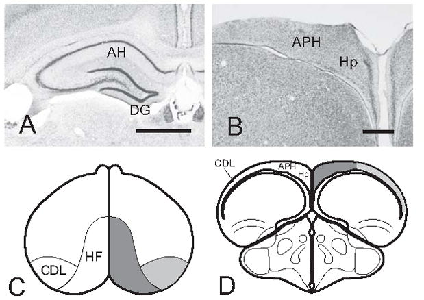
(A, B) Histological comparison of the hippocampal formation between the rat and the pigeon. (A) Layers of the dentate gyrus (DG) and cornu ammonis (CA) are conspicuous in the rat. (B) The V-shaped layer is the only readily apparent structure in the pigeon hippocampus (Hp). APH - area parahippocampalis. (C,D) Location and extent of the pigeon hippocampal formation (HF, dark gray) and dorsolateral corticoid area (CDL, light gray). (C) Dorsal view. (D) Transverse section. Scale bars in A,B = 1 mm. Reproduced with permission from Atoji & Wild (2007) following Rattenborg et al. (2010).