Figure 11.

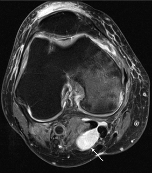
Axial magnetic resonance T2 fat sat image depicting Baker's cyst with its neck between the semimembranosus and the medial gastrocnemius tendons. Image also depicts prepatellar bursitis.

Axial magnetic resonance T2 fat sat image depicting Baker's cyst with its neck between the semimembranosus and the medial gastrocnemius tendons. Image also depicts prepatellar bursitis.