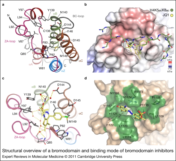
Figure 3.
Structural overview of a bromodomain and binding mode of bromodomain inhibitors. (a) Ribbon diagram of the first BRD of BRD4. The main structural elements as well as the acetyl lysine binding site residues are labelled. (b) Superimposition of a diacetylated BET substrate peptide and the inhibitor JQ1. Inhibitor and peptide molecules are shown in stick representation and are coloured according to atom types. (c) Binding of JQ1 to the bromodomain of BRD4. Conserved water molecules in the active site are highlighted and hydrogen bonds are shown as dashed lines. (d) Complex of ischemin with CREBBP (Ref. 176).

