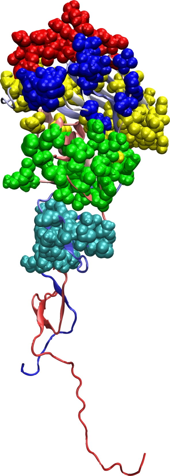
Figure 1.

The tertiary structure of the HA1 domain of H3 HA (PDB code: 1HGF). The surface of HA1 facing outward is the exposed surface when the HA trimer is formed. The other two HA1 domains (not shown) in the HA trimer are located at the back of the structure displayed here. The solid balls represent five epitopes. Colour code: blue, epitope A; red, epitope B; cyan, epitope C; yellow, epitope D; green, epitope E.
