Figure 2.
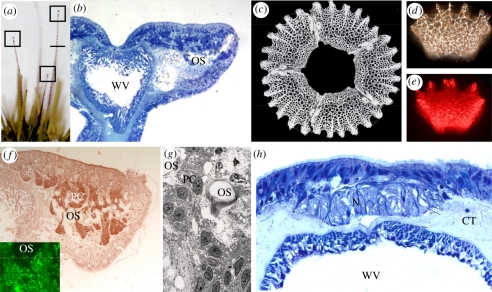
Green sea urchin tube feet. (a) Extended tube feet of a green sea urchin (maximum length of adult tube feet is 7–8 cm). Boxes indicate the distal tips shown in (b) and the line indicates the position of the cross-section shown in (h). (b) Structure of the right-hand side of the distal tip of a tube foot, showing position of ossicles (OS) seen in (c). (c) Array of five single crystalline ossicles removed from the tube foot. (d) Single ossicle under polarized light. (e) Single ossicle showing scattering of laser light directed at lower left corner. (f) Immunocytochemical localization of PAX6 protein in the pigment cells (PC) passing through perforations in the ossicles; inset shows the same PAX6 antibody conjugated to Qdots within pigment cell nuclei. (g) Electron micrograph of pigment cells passing through ossicle. (h) Cross-section at the level shown in figure 1a, indicating the nerve (N) running down one side of the tube foot (CT, connective tissue; WV, water vascular system of tube foot).

