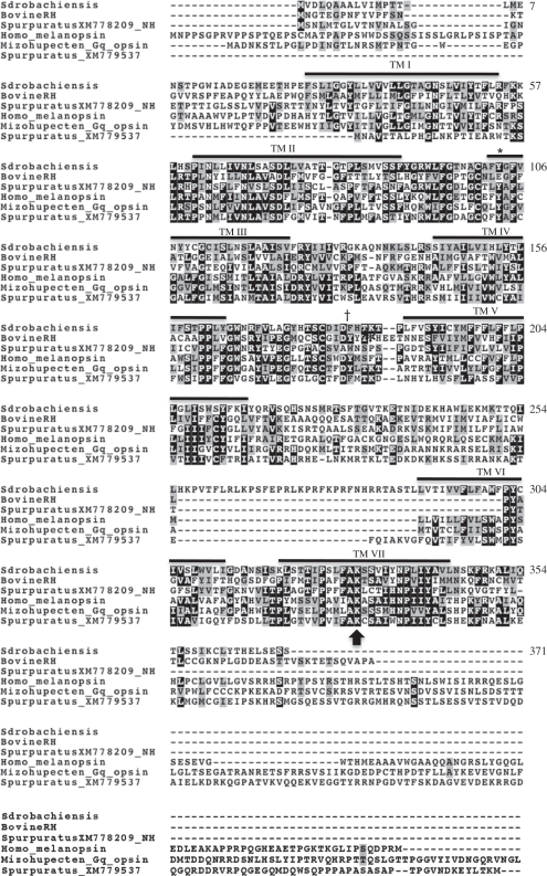
Figure 3.
Amino acid alignment of green urchin opsin and several other opsins. Identical residues between species share a black background and conserved residues share a grey background. Transmembrane domains (TM) are indicated with a solid bar, lysine attachment site for retinal is marked with an arrow, the tyrosine residue (E113/Y103) is marked with an asterisk and the aspartic acid at the proposed counterion position (D181) for the green sea urchin opsin is indicated with a dagger.

