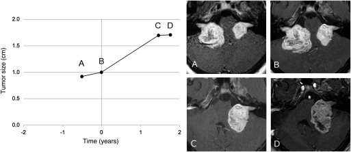
Fig. 3.
Progression of the radiological growth rate of the remaining vestibular schwannoma (VS) after resection of the contralateral tumor (Patient 9). Each point represents a magnetic resonance imaging examination (gadolinium-enhanced T1-weighted images) before and after removal of the first VS. Before surgery, the tumor grew with a radiological velocity of diametric expansion (VDE) of 4.0 mm/year. After surgery, the radiological VDE increased to 7.9 mm/year.

