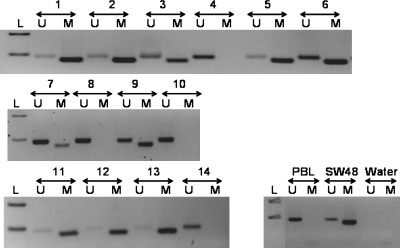
Fig. 2.
Methylation status of the MGMT promoter, as determined by methylation-specific PCR assay. DNA from normal peripheral blood lymphocytes (PBL) was used as a control for the unmethylated MGMT promoter (U), enzymatically methylated lymphocytic DNA (SW48) served as a positive control for the methylated MGMT promoter (M), and water was used as a negative control for the PCR. A 100-bp marker ladder (L) was loaded to estimate molecular size. M: PCR product amplified by methylation-specific primers; U: PCR product amplified by unmethylated-specific primers; L: ladder; SW48: methylated control DNA; PBL: unmethylated control DNA. Samples 1, 2, 3, 5, 6, 7, 9, 11, 12, and 13 were methylated with presence of band in methylated “M” lane. Band in “U” lane is because of lymphocytes and normal tissue present with tumor tissue. Cases 4, 8, 10, and 14 were unmethylated with no band in “M” lane. In control samples, PBL had a band only in “U” lane, whereas SW48 had a band only in “M” lane.

