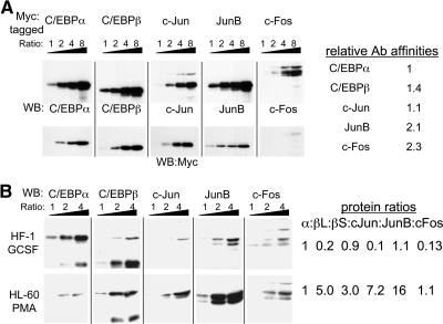
Figure 4. Semiquantitative analysis of C/EBP and AP-1 family protein levels in myeloid cells.
(A) Increasing volumes (ratio 1:2:4:8) of IVT Myc-tagged C/EBPα, C/EBPβ, c-Jun, JunB, or c-Fos were subjected to Western blotting with protein-specific antisera (upper panels) or Myc antibody (lower panels). The upper or lower sets of blots were exposed to the same secondary antibody solution for an identical time period and were subjected simultaneously to the chemiluminescence reagents and autoradiography. Relative antibody affinities were estimated by densitometric band analysis, correcting the intensities obtained with the specific antisera using the relative protein expression, as determined using the Myc antibody. (B) Nuclear extracts (2.5, 5.0, or 10.0 μg; ratio 1:2:4) from HF-1 cells cultured with G-CSF for 24 h or HL-60 cells cultured with PMA for 6 h were subjected to Western blotting with the indicated antisera. For each cell line, the series of blots were exposed to the same secondary antibody solution for an identical time period and were subjected simultaneously to the chemiluminescence reagents and autoradiography. Protein expression ratios, setting the C/EBPα (α) level to 1 in each cell line, were estimated from densitometric analysis, correcting intensities using the relative antibody affinities. βL and βS are the long and short C/EBPβ isoforms, LAP and LIP.

