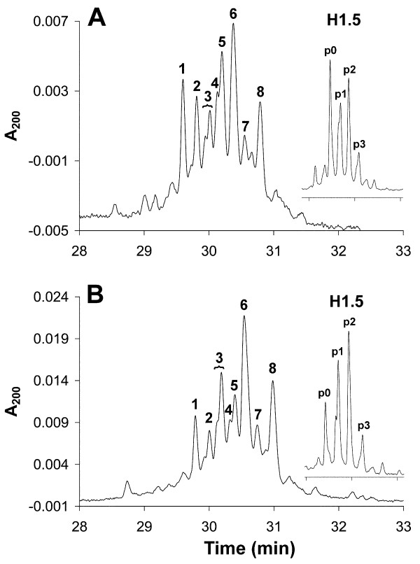
Figure 4.
High-performance capillary electrophoresis (HPCE) separations of H1 histones and reversed phase high performance liquid chromatography (RP-HPLC)-fractionated H1.5 (inset) from (A) activated T cells, and (B) exponentially growing Jurkat cells. The peaks were identified as: (1), unphosphorylated H1.5; (2) unphosphorylated H1.4; (3) monophosphorylated H1.5; (4) monophosphorylated H1.4; (5) unphosphorylated H1.3; (6) diphosphorylated H1.5, together with unphosphorylated H1.2 and possibly diphosphorylated H1.4; (7) monophosphorylated H1.3; and (8) monophosphorylated H1.2 together with triphosphorylated H1.5.

