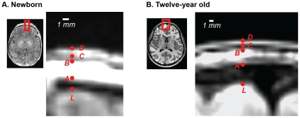
Figure 4. Components of brain-scalp distance.
A. Enlarged axial slice through a newborn infant brain (subject FT2009, age 2 days). White shows scale bar. Red letters show manually selected landmarks. L: Frontal pole landmark (outer boundary of cerebral cortex). A: Inner boundary of cranial bone marrow. B: Outer boundary of cranial bone marrow. C: Inner boundary of cutis. D: Outer boundary of cutis. B. Enlarged axial slice through a child brain (subject CBP, age 12 years). White shows scale bar. Red letters show manually selected landmarks.

