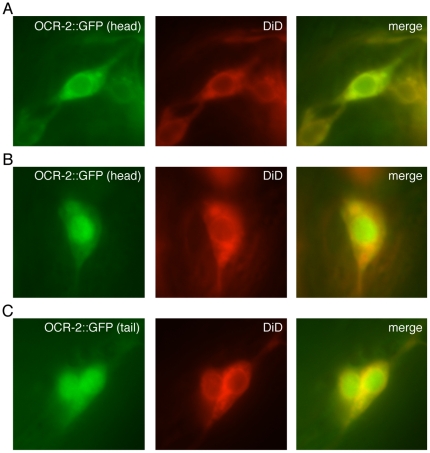
Figure 4. A GFP fusion to the carboxy-terminus of the full-length OCR-2 channel can be detected in the nucleus.
GFP was fused downstream of and in frame with full-length OCR-2 and expressed under the control of the ocr-2 promoter (ocr-2p::ocr-2::gfp). Transgenic animals were incubated with the lipophilic dye DiD (shown in red), which is taken up by several of the OCR-2 expressing neurons and was used to visualize the neuronal cell body. (A) As previously reported for an amino-terminal GFP fusion [5], flourescence was detected in the cell body and cilia of the AWA, ASH, ADL and ADF head neurons and PHA and PHB tail neurons [5]. The cell body of a head neuron is shown. (B–C) In 82/203 (40%) transgenic animals examined, distinct nuclear accumulation of GFP was detected in at least one neuron. Head (B) and tail (C) neurons are shown. Numbers represent the combined data of 3 independent transgenic lines. To show the animal-to-animal variability in subcellular localization within a transgenic line, all three images shown here were obtained from a single line.

