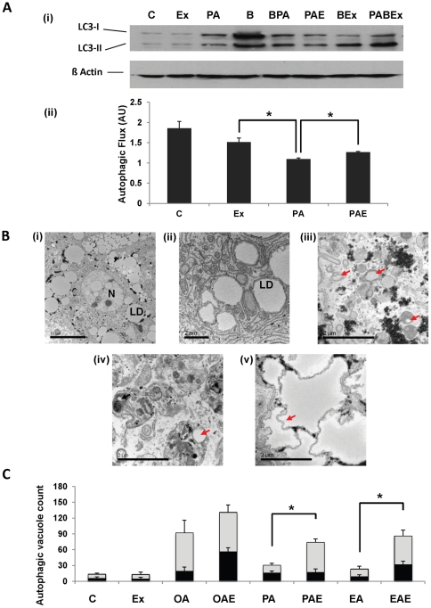
Figure 5. Exendin-4 stimulates autophagic vacuoles formation in human hepatocytes.
(A) (i) Immunoblot for LC3 in palmitic acid and/or exendin-4 treated samples in presence or absence of bafilomycin A1. (B: bafilomycin A1; BPA: bafilomycin with palmitic acid; BEx: bafilomycin with exendin-4; PABEx: palmitic acid with bafilomycin and exendin-4). (ii) Autophagic flux depicted as values obtained after taking the ratio of densitometry values of LC3-II values from bafilomycin treated samples to their respective bafilomycin-free counterparts. Note the increase in autophagic flux in response to exendin-4 in the presence of palmitic acid against palmitic acid alone. (B) Electron microscopy of primary human hepatocytes (i) showing fat loading, (N: nucleus; LD: lipid droplet), (ii) structure of lipid droplets, (iii) autophagosomes containing lipid droplets (red arrow), (iv) autophagolysosome with lipid autophagosome and lipid droplet (iv) and, (v) shriveled lipid droplet (red arrow). Magnification: (i) – (ii) x3.5E102, (iii) – (v) x25E103. (C) Quantification of autophagic vacuoles (autophagosomes containing lipid droplets – dark bands, and autophagolysosomes containing lipid droplets – light bands) by integer scoring. #p<0.001, *p<0.05, Student's t-test, three independent experiments were performed.

