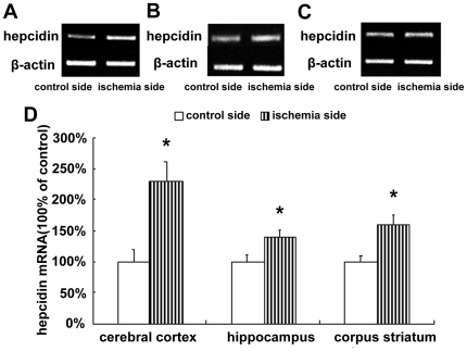
Figure 3. mRNA expression of hepcidin in ischemic brain.
Rats were subjected to 60 min MCAO and 24 hour reperfusion. RT-PCR analyses were carried out to evaluate the mRNA level of hepcidin in the cerebral cortex (A), hippocampus (B) and corpus striatum (C) of contralateral and ipsilateral side. Fig. 3D shows the summary of hepcidin expression from three animals in each group. The expression of hepcidin was significantly increased in the ischemic cerebral cortex, hippocampus and corpus striatum at 24 h. Results are presented as Mean ± SEM, n = 3. *p<0.05 v.s. contralateral side.

