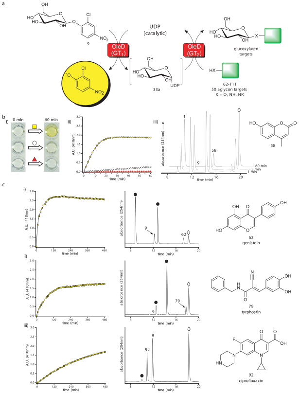
Fig. 5.
Utilizing a colorimetric screen for glycosyl transfer. (a) Scheme for colorimetric screen using the single enzyme (TDP-16) coupled format. (b) Evaluation of the colorimetric assay with 58 as the final acceptor. The reactions contained 0.5 mM 9 as donor, 0.5 mM 58 as acceptor, 5 μM UDP, and 11μM TDP-16 in a final total volume of 100 μl with Tris-HCl buffer (50 mM, pH 8.5) in a 96-well plate incubated at 25°C for one hour. (i) Qualitative color change after one hour for the full reaction (yellow square), a control lacking the final acceptor 58 (white circle), and a control lacking UDP (red triangle). (ii) Δ410 nm over one hour for the full reaction (yellow squares), a control lacking the final acceptor 58 (white circles), and a control reaction lacking UDP (red triangles). (iii) HPLC chromatograms of full reaction at 1, 5, and 60 min where 1 is desired product, 9 is the donor, 58 is the target aglycon and ⋄ represents 2-chloro-4-nitrophenolate. (c) The absorbance data and HPLC chromatograms of three representative hits [(i) 62 (genistein), (ii) 79 (tyrphostin), or (iii) 92 (ciprofloxacin)] from the broad 50 compound panel screen using the single enzyme (TDP-16) coupled format. In HPLC chromatograms 9 indicates donor; 62, 79 or 92 represent target aglycon; ⋄ indicates 2-chloro-4-nitrophenolate; and ● depicts glucosylated product(s). For the overall results of the 50 compound screen, additional representative absorbance plots and chromatograms, and combined HPLC and LC/MS characterization, see Supplementary Fig. 19–21 and Supplementary Table 6.

