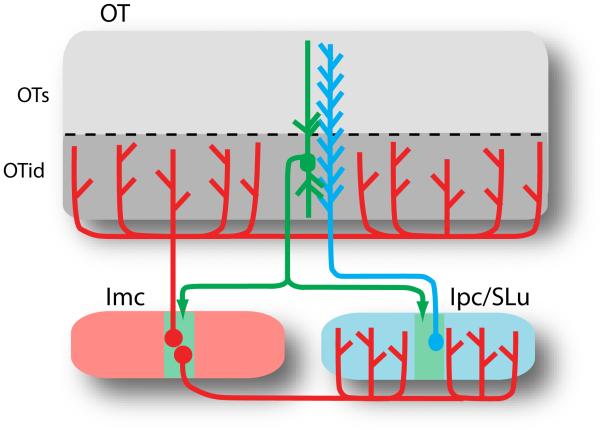
Figure 4.
The midbrain network for stimulus selection in birds. Schematic of cellular connections between the OT and the isthmic nuclei. OT neurons (green) send axons to cholinergic neurons (blue) in the Ipc and SLu and to GABAegic neurons (red) in the Imc. The OT is divided into superficial (OTs, layers 2-9) and intermediate and deep layers (OTid, layers 10-13) [5]. The green zones indicate the restricted termination zones for the OT neuron. Ipc neurons project back topographically to the OT, preferentially to the superficial layers; SLu neurons project back topographically, preferentially to the intermediate and deep layers, as well as to the thalamus and pretectum (not shown). Imc neurons project broadly to the OTid, Ipc and SLu. Abbreviations: Imc, nucleus isthmi pars magnocellularis; Ipc, nucleus isthmi pars parvocellularis; SLu, nucleus semilunaris.

