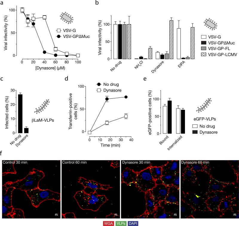
Figure 6. Dynasore reduces EBOV GPΔMuc-dependent infection by inhibiting viral uptake.
(A) Dynasore inhibits infection by VSV-GPΔMuc. Vero cells were treated with the indicated concentrations of the dynamin inhibitor dynasore and then exposed to VSV-GPΔMuc or VSV-G. Infection was scored by counting eGFP-positive cells. (B) Dynasore inhibits infection by VSV-GP-FL and VSV-GPΔMuc but not VSV-LCMV GP. Vero cells were pre-treated with 1% DMSO, 120 μM dynasore, 25 μM EIPA or 20 mM ammonium chloride and then exposed to virus. Infection was scored by counting eGFP-positive cells (VSV-GP and VSV-G) or Alexa-488-positive cells after immunostaining for the viral glycoprotein (VSV-LCMV GP). (C) Dynasore inhibits infection of βLaM-VLPs. Entry of βLaM-VLPs into Vero cells pre-treated with dynasore was quantitated by flow cytometry. (D) Dynasore inhibits transferrin uptake. Dynasore-treated cells were pulsed with 1 μg/mL Alexa-647-labeled transferrin for the indicated times, and transferrin internalization was assessed by flow cytometry. (E) Effect of dynasore on VLP uptake (flow cytometry). Dynasore-treated cells were spin-inoculated with eGFP-VLPs and internalization was allowed to occur for 30 min at 37°C. Internalized VLPs were assayed by flow cytometry. (F) Dynasore inhibits uptake of eGFP-VLPs (microscopy). Vero cells pre-treated with dynasore were spin-inoculated with eGFP-VLPs, incubated in the presence of drug at 37°C for the indicated times, and then fixed. Representative maximal Z projections of middle slices from confocal microscopy are shown. Scale bars, 5 μM. (A-E) Means +/-SD of three replicates are shown.

