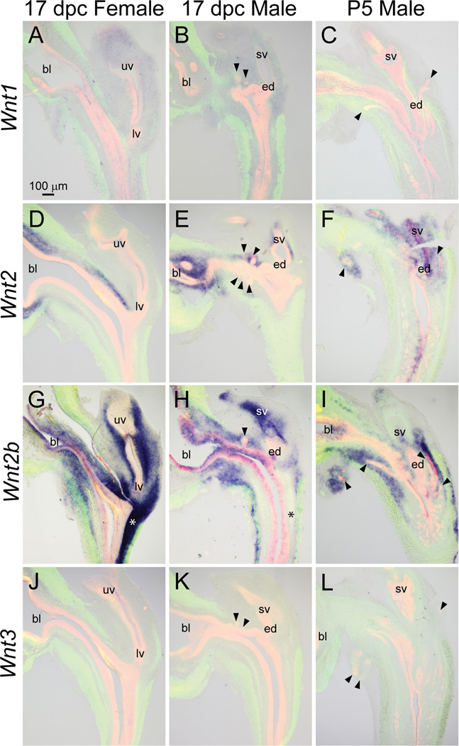
Fig. 4.
Wnt1, 2, 2b, and 3 mRNA expression patterns in developing and neonatal mouse LUT. Near mid-sagittal sections (50 µm) of 17 dpc male and female LUT and postnatal day 5 prostate were stained by ISH to visualize mRNA expression (purple) patterns of (A–C) wingless-related MMTV integration site 1 (Wnt1), (D–F) Wnt2, (G–I) Wnt2b, and (J–L) Wnt3. Sections were then stained by immunofluorescence with an anti-smooth muscle actin alpha 2 (ACTA2) antibody that recognizes muscularis mucosa and muscularis propria (green) and an anti-cadherin 1 (CDH1) antibody that recognizes all urothelium (red). Results in each panel are representative of three males and three females. Arrowheads indicate prostatic buds. Asterisks indicate regions of sexually dimorphic mRNA expression. Abbreviations used are BL: bladder, ED: ejaculatory duct, LV: lower vagina, SV: seminal vesicle, UV: upper vagina. All images are of the same magnification.

