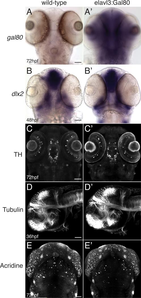
Figure 1.
Pan-neuronal expression of Gal80 does not affect CNS development. Whole-mount embryos (transgenic Tg(elavl3:Gal80)zc64 or non-transgenic sibling), ventral views (except D), anterior to the top, scale bar 50 μm. Images are confocal projections (except A and B). (A-A’) Brightfield images showing expression of gal80 by in situ in wild-type (A) and transgenic embryos (A’). (B-B’) dlx2 in situ expression in wild-type and transgenic embryos is similar. (C-C’) Pattern of tyrosine hydroxylase (TH) antibody expression is similar in wild-type and transgenic embryos. (D-D’) Axon tract architecture appears similar in wild-type and transgenic embryos (lateral views, anterior to the left). (E-E’) Acridine orange staining for apoptotic cells is similar in wild-type and transgenic embryos.

