Figure 2.
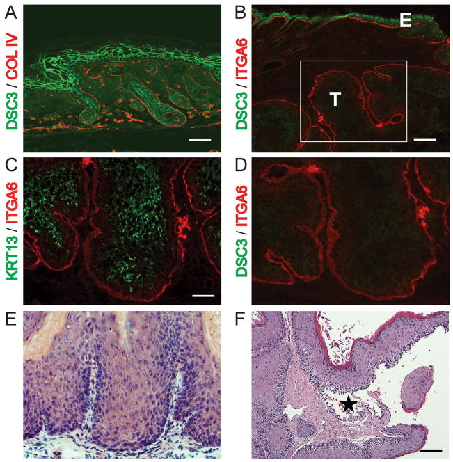
Tumor induction and Dsc3 ablation in mutant mice. Immunofluorescence microscopy (A-D) using antibodies against DSC3, keratin 13 (KRT13) and the basement membrane markers α6 integrin (ITGA6) and collagen IV (COLIV). (A) DSC3 (green) and COLIV (red) staining of newborn epidermis. Bar, 50μm (B) DSC3 (green) and ITGA6 (red) staining of a tumor from a RU486-treated mutant mouse. Note the strong DSC3 staining in the epidermis (E) and the absence of DSC3 staining in the tumor tissue (T) that has penetrated the dermis. Bar, 100μm (C) KRT13, an early marker for tumor expression [20], was frequently induced in DSC3-negative tumor tissues (Bar, 50μm). (D) DSC3 staining of the section shown in (C) and in the boxed area of panel (B). Note the complete absence of DSC3 staining in the tumor cells. (E) Histological staining of the tumor section shown in (B and C). (F) Histological section of a mutant tumor. Note the suprabasal acantholysis (*) in the tumor tissue. Bar, 100μm

