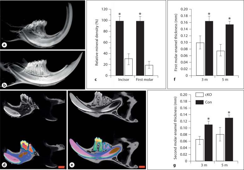
Fig. 2.
X-ray and micro-CT of teeth. X-ray analysis of the enamel in the Bmp2-cKO (a) and control (b) mice. The mineral density of incisors and molars from Bmp2-cKO mice was decreased compared to control mice (c). The mineral density of teeth from the control mice acts as 100%, and asterisks (∗) show significant differences between the control and Bmp2-cKO mice. The enamel layer was thin and the mineral density decreased in the 3-month-old Bmp2-null mice. Micro-CT analysis of teeth of the Bmp2-cKO (d) and control (e) mice. Light blue = Enamel thickness; yellow = mantle dentin; green = root dentin; red = dental pulp chamber; blue = alveolar bone. The mandibles from 3-month-old mice were subjected to micro-CT analysis by Numira, Inc. as described in Materials and Methods using parameters that were developed to measure the enamel width of the first and second molars. Significance was different between the control and Bmp2-cKO groups (f, g) (p < 0.05). The mineral density of the enamel from the null mice was reduced as compared to the control groups. m = Months. Scale bar = 1 mm.

