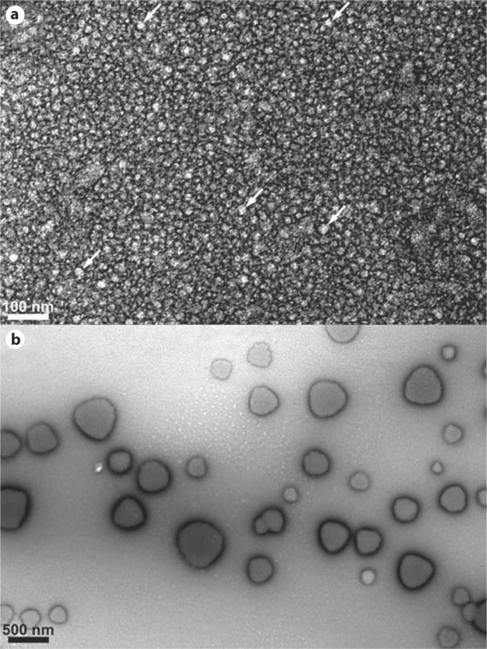
Fig. 1.

TEM images showing the T21I mutant at pH 5.8, 0.4 mg/ml, and 37°C (a). The particles were 18.2 nm in diameter on average, which is in approximate agreement with DLS. White arrows highlight 5 of the most clearly apparent oligomers. b The P41T mutant formed large aggregates at 37°C with diameters ranging from 100 to 500 nm, again in agreement with DLS. Scale bars = 100 and 500 nm, respectively.
