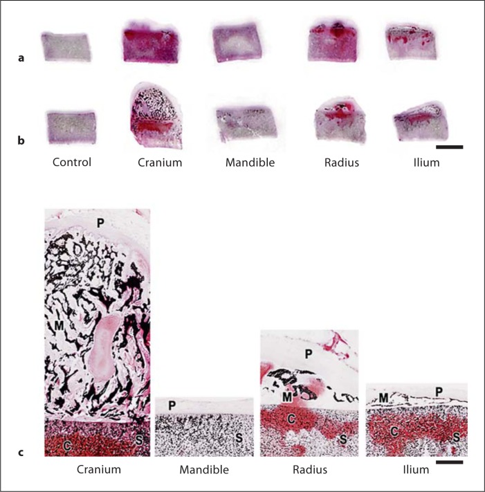
Fig. 2.
Representative thick (5 μm) sections from ∼5-month-old calf constructs retrieved after 10 (a) or 20 (b) weeks of implantation in nude mice. Sections in b were obtained from the samples in figure 1a. Two examples of sections from control scaffolds without sutured periosteum are also shown. Sections were treated with von Kossa and safranin-O red solutions for the detection of phosphate and proteoglycans, respectively. Over the two time intervals for implantation, the size or extent of development differed among the constructs sutured with periosteum, as well as the presence and distribution of phosphate (as a surrogate for mineral deposition) and proteoglycans. Section enlargements of 20-week implants show details of structure and staining (c). S = Scaffolds; C = cartilage proteoglycans; M = mineral; P = periosteum. Scale bars = 5 mm (a, b) and 1 mm (c).

