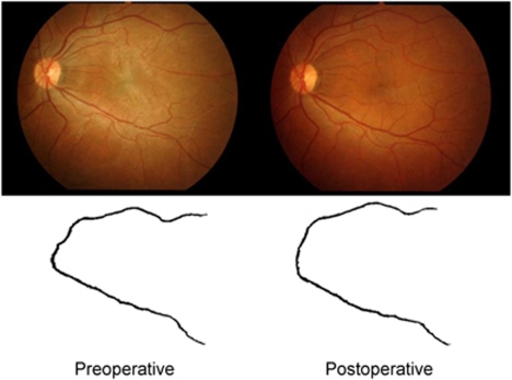
Figure 1.
An example of a preoperative and postoperative image pair from a patient with an epiretinal membrane. A patient's fundus photographs are shown (top left) preoperatively with an epiretinal membrane and (top right) postoperatively. The binary images of major vessels from (bottom left) preoperative and (bottom right) postoperative fundus photographs are shown.

