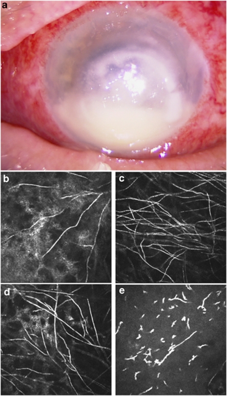
Figure 1.
(a) Photography of the left eye showing a large corneal central whitish irregular infiltrate with 2 mm hypopion. (b–d) In vivo confocal microscopy images (400 × 400 μm2) showing numerous hyper-reflective interlocking and branching linear structures, 5–7 μm width and 200–400 μm length, possibly corresponding to fungal hyphae. These images were obtained in the stroma at the boundaries of the central corneal infiltrate: (b) in the nasal area, depth: 115 μm; (c) in the superior area, depth: 223 μm; (d) in the superior area, depth: 106 μm. (e) In vivo confocal microscopy image (400 × 400 μm2) of the corneal stroma (45 μm depth) showing a stromal oedema with infiltration of inflammatory cells. The stroma appeared as a dark background with hyper-reflective small structures with an occasional dendritic-like pattern corresponding to inflammatory cells.

