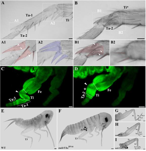
Fig. 3.
nub and Ubx are required for the proper leg formation in Acheta. (A-B) Magnified images of the metathoracic tibia and tarsus of wild type and class I first nymphs, respectively. (A1-A2) Close-up views of the boundaries between the first and second tarsomeres (A1) and between the tibia and first tarsomere (A2) in wild type. In order to better visualize their morphologies, spines and spurs were artificially colored. Ta-1/Ta-2 boundary bears a pair of spines and two spurs covered with bristles (arrowheads), while four spurs free of bristles surround Ti/Ta-1 boundary. (B1-B2) Close-up views of the corresponding structures in nub-depleted individuals. The distal region of the tibia now bears one pair of spurs covered with bristles (B1, arrowheads) and one pair of spines (B2), both cuticular features that are normally found on the first tarsomere. (C, D) Ubx expression patterns in the hind legs of wild type and nub-depleted embryos, respectively. (C) The T3 legs of wild type Acheta embryos exhibit an ectodermal Ubx expression in the tibia and mesodermal signal in the femur, first and second tarsomeres. (D) In contrast, nub RNAi embryos have a novel mesodermal expression of Ubx in the mid-distal region of the tibia (asterisk) and a single ring in the tarsus that corresponds to the second tarsomere. In both panels, an arrowhead indicates the absence of signal in the small area of the proximal tibia. (E, F) Lateral views of wild type and nub/Ubx RNAi embryos, respectively. The latter grows ectopic appendages on the first abdominal segment and has severely shortened thoracic legs. (G-I) Dissected hind legs of wild type (G), nub RNAi (H), and nub/Ubx RNAi embryos (I). Compared to wild type and nub RNAi alone, the T3 legs of double nub/Ubx RNAi embryos are more drastically reduced in size due to a significant shortening of the femur, tibia, and tarsus. White solid lines mark the boundaries between individual segments. Scale bars: 50 μm (A, B, C, and D); 100 μm (E-I). Abbreviations: WT, wild type; T3, hind leg; Fe, femur; Ti, tibia; Ta, tarsus; Ta 1-3, tarsomeres 1-3; Ti*, fused tibia and first tarsomere; A1, first abdominal segment.

