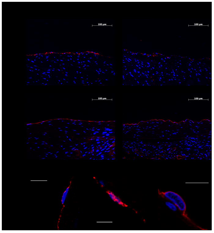
Figure 2. Representative immunofluorescence and confocal images of the RAA and LAA expression of thrombomodulin and EPCR.

(A) Representative captures of thrombomodulin and EPCR on the endocardium of the RAA and LAA. Thrombomodulin and EPCR are red-stained. Blue DAPI was used to stain the nuclei. (B) Confocal microscopy captures of thrombomodulin (left), von Willebrand factor (middle) and EPCR (right), all red-colored, in representative endothelial cells. Von Willebrand factor staining was used as an endothelial positive control. Blue Topro-3 was used to stain the nuclei. The scale bars indicate 10 μm.
