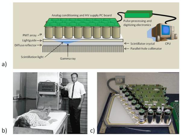
Figure 3.
a) The basic structure of the Anger Camera comprises a collimator, a monolithic scintillator crystal, a light guide that allows light to spread, and an array of photomultiplier tubes (PMTs) with related electronics. Position estimation was originally performed with analog circuitry; in current systems PMT outputs are digitized and all processing is digital. b) Hal Anger shown with early example of his camera being applied in a clinical setting (Reprinted by permission of the Society of Nuclear Medicine from: Nuclear Medicine Pioneer, Hal O. Anger, 1920–2005. J Nucl Med Technol. 2005; 33(4): 250-253). c) A cutaway of an actual camera (Courtesy of M. Wernick and J. Aarsvold).

