FIG. 1.
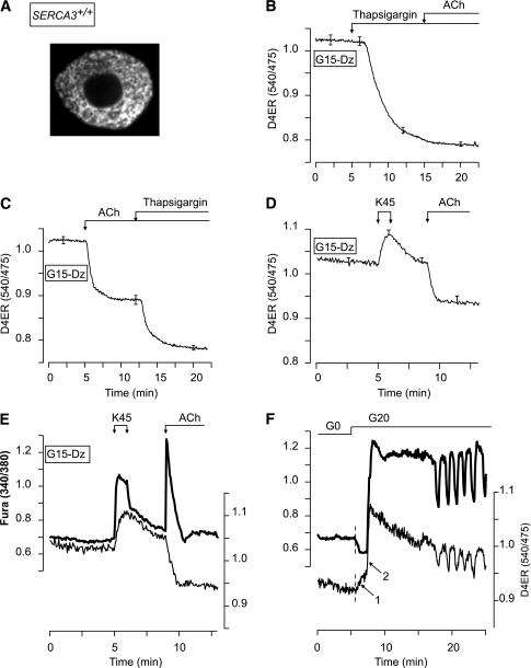
Validation of D4ER as a reporter of [Ca2+]ER changes and of combined measurements of [Ca2+]c and [Ca2+]ER. A: Confocal image of a single β-cell expressing D4ER. B–D: β-Cell [Ca2+]ER measurements. Cells were perifused with 15 mmol/L glucose (G15) in the presence of 250 µmol/L of the KATP channel opener Dz. As indicated, 1 µmol/L thapsigargin, 100 µmol/L ACh, or 45 mmol/L KCl (K45) was added. E and F: Simultaneous measurement of [Ca2+]c (FuraPE3) and [Ca2+]ER (D4ER) in β-cells. E: The perifusion medium contained 15 mmol/L glucose (G15) and 250 µmol/L Dz throughout. β-Cells were stimulated with 45 mmol/L KCl (K45) and 100 µmol/L ACh as indicated. F: β-Cells were perifused in glucose-free medium (G0) and then stimulated with 20 mmol/L glucose (G20). B–D: Means ± SE for 23–44 cells from three to four experiments with three islet preparations. E and F: Representative traces from 9 to 42 cells from three experiments with three islet preparations.

