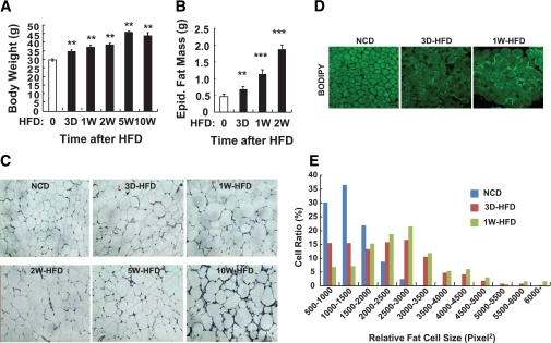
FIG. 1.
In 3 days of HFD, body weight, fat mass, and adipocyte size are increased significantly. Eight-week-old C57BL6J male mice were fed NCD until they were subjected to 60% HFD. HFD was treated to the mice 0 (NCD), 3 days (3 D), 1 week (1 W), 2 weeks (2 W), 5 weeks (5 W), or 10 weeks (10 W) before death. At the age of 18 weeks, all mice were killed and subjected to several analyses. A: Body weight of the mice at the end of the experiment. n = 8 at each time point. Increase of body weight started to be observed by 3 days of HFD. Body weight of mice with 5-week HFD and 10-week HFD was not significantly different. **P < 0.05. B: Epididymal (Epid.) fat mass. n = 8 at each time point. **P < 0.05; ***P < 0.001. C: Histology analysis of epididymal adipose tissue. The epididymal adipose tissues were subjected to hematoxylin-eosin staining. n = 4 at each time point. D and E: Further qualitative changes of adipocyte size and morphology by short-term HFD were assessed by histology analysis of whole-mount epididymal adipose tissue. D: BODIPY (boron-dipyrromethene) staining of whole-mount adipose tissue from the mice treated with NCD or HFD. Adipocyte size was markedly increased by 3 days of HFD. n = 6. Signal intensity of BODIPY staining was adjusted for optimal measurement of adipocyte size using ImageJ software. E: Distribution of adipocyte size in epididymal adipose tissue from mice treated with NCD or HFD. (A high-quality digital representation of this figure is available in the online issue.)

