
Figure 5.
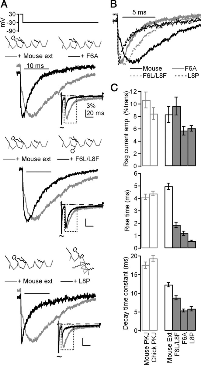
The identities and orientation of a phenylalanine and two lysine residues are key determinants of resurgent current kinetics. A, Mean currents evoked by the resurgent current protocol in CA3 neurons with control mouse (gray) and mutant (black) β4 peptides, normalized to peak current evoked by repolarization. Top, F6A, n = 7; middle, F6A/L8F, n = 9; bottom, L8P, n = 9. Insets, Mean currents normalized to peak transient current at +30 mV. Peptide schematics were rendered in Deepview (Swiss Institute of Bioinformatics). Gray indicates α carbon trace; black indicates side chains of K2, F6, K9, K10, and K16. Other side chains are blanked for clarity. B, Overlay of mean currents from A, normalized to resurgent current peak. C, Top, Mean amplitude of currents evoked at −30 mV after 10 ms conditioning step to +30 mV, normalized to transient current at +30 mV. Middle, Mean rise time of resurgent currents evoked at −30 mV. Bottom, Mean decay time constants (τ) of resurgent currents evoked at −30 mV.