
Figure 7.
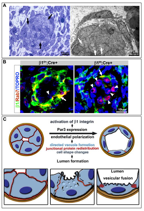
Arrest of lumen formation and excess of cytosolic vacuoles in β1 deleted endothelial cells (ECs). (A) Semithin (left) and EM (right) analysis of luminal occlusion in E15.5 β1f/f;Cre+ animals demonstrates accumulation of multiple vesicles/vacuoles within the cell cytoplasm (arrows). (B) Evaluation of Rab7 (red) in context of β1 integrin (green) demonstrates Rab7 in the basal aspect of the endothelium and at the smooth muscle cell (SMC)/ EC junction with β1 integrin (yellow), but minimal expression within the endothelial layer (arrowhead). Upon β1 deletion, a dramatic increase of Rab7 expression in β1 ablated ECs was noted (right panel, arrowheads) with some maintenance of co-expression with β1 integrin in the SMC layer (arrow). (C) Schema depicts a working model of the cascade of events that drive lumen formation. Activation of β1 integrin instructs Par3 expression which then allows for polarization of the endothelium. Vesicular fusion to the apical cell membrane results in redistribution of junctional and adhesion proteins (in red, arrows), change in cell shape from cuboidal to squamous, and acquisition of a vascular lumen.